 Indietro
Tissues: Structure, Function, and Classification in Human Anatomy & Physiology
Guida di studio - Note intelligenti
 Appunti personalizzati basati sui tuoi materiali, ampliati con definizioni chiave, esempi e contesto.
Tissues: An Overview
Definition and Types of Tissues
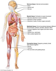
Tissues are groups of cells that are similar in structure and perform a common function. The human body contains four primary tissue types:
Epithelial tissue – covers surfaces and lines cavities
Connective tissue – supports and binds other tissues
Muscle tissue – responsible for movement
Nervous tissue – controls and communicates
Preparation of Tissue for Microscopy
Steps in Tissue Preparation
To study tissues under a microscope, samples must be properly prepared. The main steps include:
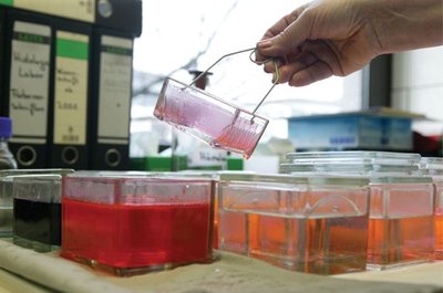
Fixation – preserves tissue structure
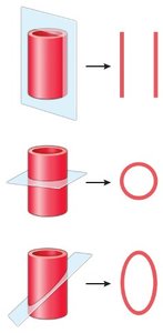
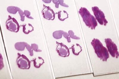
Sectioning – slices tissue into thin sections
Staining – enhances contrast for visualization
Epithelial Tissue
Characteristics and Functions
Epithelial tissue consists of sheets of cells that cover body surfaces or line body cavities. It serves several functions:
Protection
Absorption
Filtration
Excretion
Secretion
Sensory reception
Key traits include polarity (apical and basal surfaces), specialized contacts, support by connective tissue (basement membrane), avascularity but innervation, and high regenerative capacity.
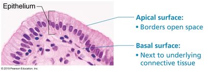
Classification of Epithelium
Epithelial tissues are classified by:
Number of cell layers: Simple (one layer) or Stratified (multiple layers)
Cell shape: Squamous (flat), Cuboidal (cube-like), Columnar (tall)
Stratified epithelia are named according to the shape of cells in the apical layer.
Types of Simple Epithelia
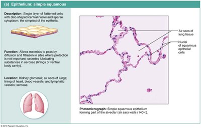
Simple Squamous Epithelium: Filtration and rapid diffusion; found in kidney glomeruli, alveoli of lungs, endothelium, and mesothelium.
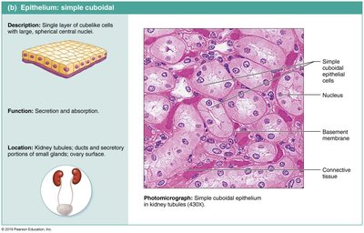
Simple Cuboidal Epithelium: Secretion and absorption; found in kidney tubules and ducts of glands.
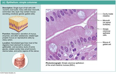
Simple Columnar Epithelium: Absorption and secretion; lines most of the digestive tract.
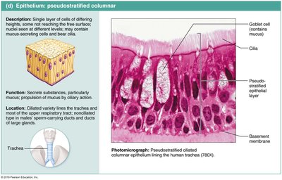
Pseudostratified Columnar Epithelium: Secretion and movement; ciliated type lines trachea and upper respiratory tract.
Types of Stratified Epithelia
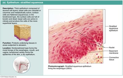
Stratified Squamous Epithelium: Protection; forms the outer layer of skin and lines body orifices.
Stratified Cuboidal & Columnar Epithelium: Rare; found in ducts of larger glands and parts of the pharynx and male urethra.
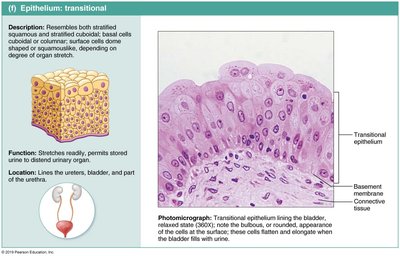
Transitional Epithelium: Stretches to allow expansion; lines ureters, bladder, and part of urethra.
Glandular Epithelium
Glands and Their Classification
A gland is one or more cells that make and secrete a particular product (secretion). Glands are classified by:
Where product is released: Endocrine (internal) or Exocrine (external)
Number of cells: Unicellular or Multicellular
Endocrine vs. Exocrine Glands
Endocrine glands: Ductless, secrete hormones directly into bloodstream; most are multicellular.
Exocrine glands: Secrete products onto body surfaces or into cavities via ducts; can be unicellular (e.g., goblet cells) or multicellular.
Unicellular Exocrine Glands
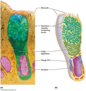
Unicellular exocrine glands, such as goblet cells, are found in epithelial linings of intestinal and respiratory tracts. They produce mucin, which forms mucus.
Modes of Secretion
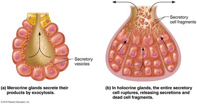
Merocrine: Secrete products by exocytosis (e.g., sweat glands, salivary glands)
Holocrine: Accumulate product until cell ruptures (e.g., sebaceous glands)
Connective Tissue
Characteristics and Functions
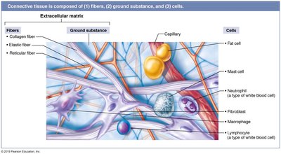
Connective tissue is the most abundant tissue in the body. It binds, supports, protects, insulates, stores fuel, and transports substances. All connective tissues share three main components:
Ground substance – unstructured material filling space between cells
Fibers – provide support (collagen, elastic, reticular)
Cells – various types depending on tissue
Types of Connective Tissue
Connective tissue proper (loose and dense types)
Cartilage (hyaline, elastic, fibrocartilage)
Bone (osseous tissue)
Blood
Loose Connective Tissue: Areolar
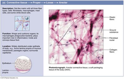
Areolar connective tissue is the most widely distributed CT in the body. It supports and binds other tissues, holds body fluids, defends against infection, and stores nutrients.
Loose Connective Tissue: Adipose
Adipose tissue is specialized for nutrient storage, insulation, and shock absorption. Adipocytes make up 90% of the tissue.

Loose Connective Tissue: Reticular
Reticular connective tissue forms a delicate network (stroma) in lymph nodes, spleen, and bone marrow.
Dense Connective Tissue: Regular
Dense regular connective tissue contains parallel bundles of collagen fibers, providing strength in one direction. Found in tendons and ligaments.
Dense Connective Tissue: Irregular
Dense irregular connective tissue has thicker collagen bundles arranged irregularly, providing strength in multiple directions. Found in fibrous capsules and dermis.
Dense Connective Tissue: Elastic
Elastic connective tissue contains many elastic fibers, allowing stretch and recoil. Found in ligaments of vertebral column and walls of large arteries.
Cartilage
Cartilage is tough but flexible, avascular, and lacks nerve fibers. Types include:
Hyaline cartilage: Most abundant; provides support with pliability. Found in articular cartilage, costal cartilage, nose, trachea, and embryonic skeleton.
Elastic cartilage: Contains more elastic fibers; found in external ear and epiglottis.
Fibrocartilage: Resists compression and tension; found in intervertebral discs and knee cartilage.
Bone (Osseous Tissue)
Bone tissue is rigid due to inorganic calcium salts. Osteoblasts produce matrix; osteocytes reside in lacunae. Osteons are the structural unit. Functions include support, protection, fat storage, and hematopoiesis.
Blood
Blood is an atypical connective tissue consisting of cells (erythrocytes, leukocytes, platelets) in a fluid matrix (plasma). It transports nutrients, wastes, and gases.
Muscle Tissue
Types and Functions
Muscle tissue is responsible for movement and is highly vascularized. It contains myofilaments (actin and myosin) for contraction. Types include:
Skeletal muscle: Voluntary, striated, long cylindrical cells with multiple nuclei; attached to bones or skin.
Cardiac muscle: Involuntary, striated, branching cells with central nucleus; found only in heart, connected by intercalated discs.
Smooth muscle: Involuntary, non-striated, spindle-shaped cells with central nucleus; found in walls of hollow organs.
Nervous Tissue
Structure and Function
Nervous tissue is the main component of the nervous system (brain, spinal cord, nerves). It regulates and controls body functions. Two major cell types:
Neurons: Specialized for generating and conducting nerve impulses; have dendrites (receive stimuli) and axons (transmit impulses).
Supporting cells: Non-conducting; support, insulate, and protect neurons.
