 Indietro
Tissues: Structure, Function, and Classification in Human Anatomy
Guida di studio - Note intelligenti
 Appunti personalizzati basati sui tuoi materiali, ampliati con definizioni chiave, esempi e contesto.
Tissues: An Introduction
Tissues are groups of similar cells that work together to perform specific functions in the body. Understanding the structure and function of tissues is fundamental to the study of Anatomy & Physiology, as tissues form the basis for organs and organ systems.
The Four Major Types of Tissue
Epithelial Tissue: Covers body surfaces, lines cavities, and forms glands.
Connective Tissue: Supports, binds, and protects other tissues and organs.
Muscle Tissue: Responsible for movement.
Nervous Tissue: Initiates and transmits electrical impulses.
Epithelial Tissue
General Characteristics
Covers all body surfaces, both internal and external.
Main tissue of glands.
Attached to underlying connective tissue at the basement membrane.
Lacks blood vessels (avascular); nutrients diffuse from underlying tissues.
Cells reproduce rapidly, aiding in healing and regeneration (but also scarring).
Cells are tightly packed together, forming protective barriers.
Functions: Protection, secretion, absorption, excretion, and sensation.
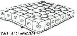
Classification of Epithelial Tissue
By Layers:
Simple: Single layer of cells.
Stratified: Multiple layers of cells.
By Shape:
Squamous: Flat and thin.
Cuboidal: Cube-shaped.
Columnar: Tall and column-like.
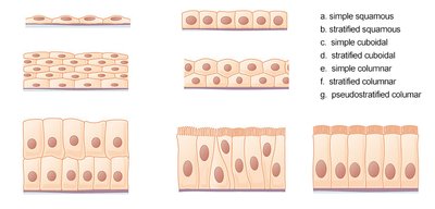
Types of Epithelial Tissue
Simple Squamous Epithelium:
Form: Single layer of flat cells.
Function: Diffusion and filtration.
Location: Air sacs of lungs, capillaries.
Simple Cuboidal Epithelium:
Form: Single layer of cube-shaped cells.
Function: Secretion and absorption.
Location: Kidney tubules, surface of ovaries.
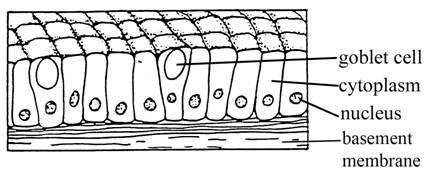
Simple Columnar Epithelium:
Form: Single layer of tall, column-like cells.
Function: Secretion and absorption.
Location: Digestive tract, uterus.
Special Features: Contains goblet cells (secrete mucus) and microvilli (increase surface area).
Stratified Squamous Epithelium:
Form: Multiple layers of flat cells.
Function: Protection.
Location: Skin, mouth, esophagus.
Pseudostratified Columnar Epithelium:
Form: Appears layered due to varying positions of nuclei, but is a single layer.
Function: Secretion and movement of mucus (often ciliated).
Location: Lining of respiratory passages, reproductive tubes.
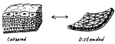
Transitional Epithelium:
Form: Multiple layers of cuboidal cells; can stretch and change shape.
Function: Allows for distension and contraction.
Location: Urinary bladder.
Glandular Epithelium:
Specialized for secretion.
Forms exocrine (e.g., sweat, salivary) and endocrine (e.g., thyroid, adrenal) glands.
Connective Tissue
General Characteristics
Most abundant tissue type in the body.
Binds structures, provides support and protection, fills spaces, stores fat, produces blood cells, and fights infection.
Composed of scattered cells within an extracellular matrix (ground substance + fibers).
Usually has a good blood supply (except cartilage and dense connective tissue).
Main Cell Types: Fibroblasts (produce fibers), macrophages (phagocytosis), mast cells (inflammation and allergy response).
Main Fiber Types: Collagenous (strong, flexible), elastic (stretchy), reticular (supportive networks).
Types of Connective Tissue
Loose (Areolar) Connective Tissue: Binds skin to underlying organs; forms thin membranes throughout the body.
Adipose Tissue (Fat): Stores energy, insulates, and cushions organs; found beneath the skin.
Dense (Fibrous) Connective Tissue: Contains thick collagenous and elastic fibers; forms tendons (muscle to bone) and ligaments (bone to bone); slow to heal due to poor blood supply.
Cartilage: Cells called chondrocytes; types include hyaline (ends of bones, nose), elastic (external ear), and fibrocartilage (intervertebral discs, knee meniscus).
Bone (Osseous) Tissue: Rigid due to mineral salts; supports and protects body structures.
Blood: Fluid connective tissue; transports substances throughout the body.
Muscle Tissue
Types and Functions
Skeletal Muscle: Voluntary, striated; attached to bones for movement.
Smooth Muscle: Involuntary, non-striated; found in walls of hollow organs (stomach, intestines, blood vessels).
Cardiac Muscle: Involuntary, striated; found only in the heart wall.
Nervous Tissue
Structure and Function
Found in the brain, spinal cord, and nerves.
Neurons: Transmit electrical signals.
Neuroglia: Support, protect, and nourish neurons.
Skin Structure and Function
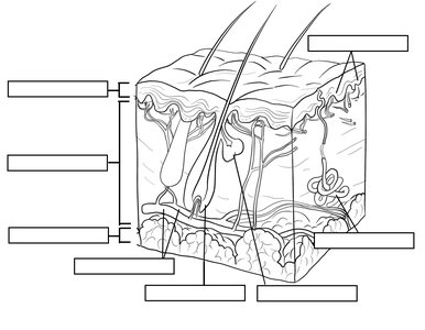
The skin is composed of multiple layers, including the epidermis (outer), dermis (middle), and subcutaneous (fatty) tissue. It contains structures such as blood vessels, sweat glands, oil glands, hair follicles, and melanocytes (pigment cells).
Special Topics
Collagen and Elastin: Key connective tissue fibers; loss leads to aging and decreased skin elasticity.
Skin Color: Determined by melanocytes and influenced by genetics and evolution (see Fitzpatrick Skin Type Scale).
Epidermolysis Bullosa: Genetic disorder affecting collagen, causing fragile skin and blistering; illustrates the importance of connective tissue in anchoring the epidermis.
Review Table: Types of Epithelial Tissue
Type | Form | Function | Location |
|---|---|---|---|
Simple Squamous | Single, flat layer | Diffusion, filtration | Lungs, capillaries |
Simple Cuboidal | Single, cube-shaped | Secretion, absorption | Kidney tubules, glands |
Simple Columnar | Single, tall cells | Secretion, absorption | Digestive tract, uterus |
Stratified Squamous | Multiple, flat layers | Protection | Skin, mouth, esophagus |
Pseudostratified Columnar | Single, appears layered | Secretion, movement | Respiratory tract |
Transitional | Multiple, stretchable | Distension | Urinary bladder |
Practice and Application
Identify tissue types from diagrams and histology slides.
Label structures in diagrams of skin and connective tissue matrices.
Compare and contrast tissue types based on structure, function, and location.
Key Terms and Definitions
Basement Membrane: Thin, non-cellular layer anchoring epithelium to connective tissue.
Matrix: Extracellular material in connective tissue, composed of ground substance and fibers.
Goblet Cell: Specialized epithelial cell that secretes mucus.
Microvilli: Small projections that increase surface area for absorption.
Cilia: Hair-like projections that move substances across cell surfaces.
Summary
Understanding the structure and function of the four major tissue types is essential for comprehending how the human body is organized and how it functions. Mastery of tissue classification, identification, and their roles in health and disease forms the foundation for further study in Anatomy & Physiology.
