 Indietro
Urinary and Reproductive Systems: Fetal Pig and Human Anatomy Study Guide
Guida di studio - Note intelligenti
 Appunti personalizzati basati sui tuoi materiali, ampliati con definizioni chiave, esempi e contesto.
Urinary System Anatomy
Overview of the Urinary System
The urinary system is responsible for filtering blood, removing waste, and regulating fluid and electrolyte balance. The main organs include the kidneys, ureters, urinary bladder, and urethra.
Kidneys: Bean-shaped organs that filter blood and produce urine.
Ureters: Tubes that transport urine from the kidneys to the urinary bladder.
Urinary Bladder: A muscular sac that stores urine until excretion.
Urethra: The tube through which urine exits the body.
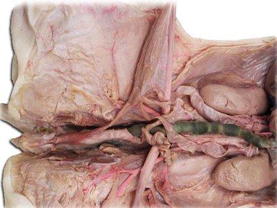
Renal Blood Supply
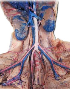
The kidneys receive blood through the renal arteries and drain blood via the renal veins. The renal artery branches from the abdominal aorta, while the renal vein drains into the inferior vena cava.
Renal Artery: Supplies oxygenated blood to the kidney.
Renal Vein: Returns filtered blood to the systemic circulation.
Segmental, Interlobar, and Arcuate Vessels: These vessels further branch within the kidney to supply and drain different regions.
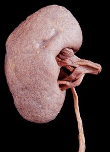
Gross Structure of the Kidney
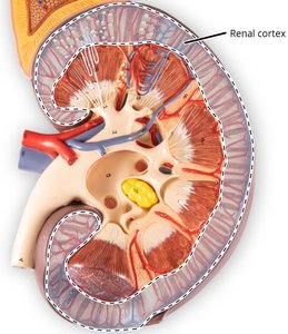
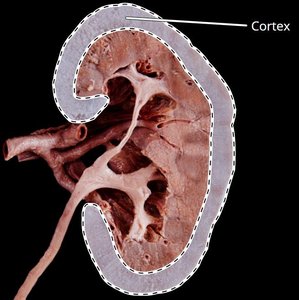
The kidney is covered by a fibrous capsule and consists of two main regions: the cortex (outer layer) and the medulla (inner region). The medulla contains renal pyramids, which are separated by renal columns.
Renal Cortex: The outer region containing glomeruli and convoluted tubules.
Renal Medulla: Contains renal pyramids, which house the loops of Henle and collecting ducts.
Renal Columns: Extensions of cortical tissue between the pyramids.
Internal Kidney Structures
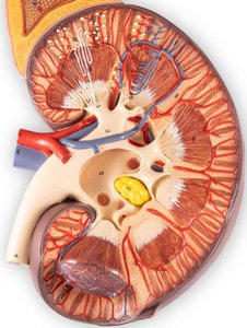
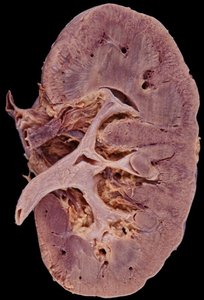
Within the medulla, the renal pyramids drain urine into minor calyces, which merge to form major calyces. These then empty into the renal pelvis, which leads to the ureter.
Renal Pyramid: Cone-shaped tissue masses in the medulla.
Renal Papilla: Tip of the pyramid where urine is discharged into the minor calyx.
Minor Calyx: Collects urine from a single pyramid.
Major Calyx: Formed by the convergence of minor calyces.
Renal Pelvis: Funnel-shaped structure that collects urine from major calyces and channels it to the ureter.
Reproductive System Anatomy
Female Reproductive Anatomy
The female reproductive system includes the ovaries, uterine tubes (fallopian tubes), uterus, and associated structures. In the fetal pig, the uterine horns are prominent, reflecting the species' reproductive strategy.
Ovaries: Produce eggs (ova) and hormones such as estrogen and progesterone.
Uterine Tubes: Transport ova from the ovaries to the uterus; site of fertilization.
Uterus: Site of fetal development; in pigs, the uterus has prominent horns.
Male Reproductive Anatomy
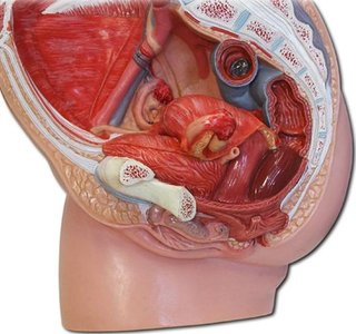
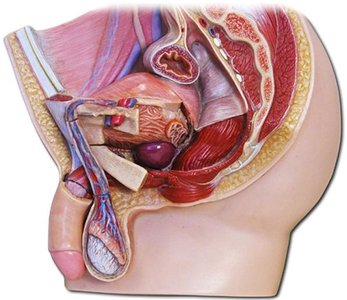
The male reproductive system consists of the testes, epididymis, ductus deferens, and penis. These structures are responsible for the production, maturation, and delivery of sperm.
Testes: Produce sperm and testosterone.
Epididymis: Site of sperm maturation and storage.
Ductus Deferens (Vas Deferens): Transports sperm from the epididymis to the urethra.
Penis: Organ of copulation and urination.
Summary Table: Major Urinary and Reproductive Structures
Structure | Function | Location |
|---|---|---|
Kidney | Filters blood, forms urine | Retroperitoneal, posterior abdominal wall |
Ureter | Transports urine to bladder | From renal pelvis to bladder |
Urinary Bladder | Stores urine | Pelvic cavity |
Urethra | Conducts urine out of body | From bladder to external opening |
Ovary | Produces ova and hormones | Pelvic cavity (female) |
Uterus | Site of fetal development | Pelvic cavity (female) |
Testes | Produce sperm and testosterone | Scrotum (male) |
Epididymis | Sperm maturation and storage | On posterior testis (male) |
Ductus Deferens | Transports sperm | From epididymis to urethra (male) |
Additional info:
The fetal pig is a common specimen for anatomical study due to its similarity to human organ systems.
In humans, the uterus is a single organ, while in pigs it is bicornuate (with two horns).
The renal cortex contains the majority of nephrons, the functional units of the kidney.
