 Indietro
Urinary System and Fluid & Acid-Base Balance: Study Guide
Guida di studio - Note intelligenti
 Appunti personalizzati basati sui tuoi materiali, ampliati con definizioni chiave, esempi e contesto.
Urinary System Anatomy & Physiology
Overview of the Urinary System
The urinary system is essential for maintaining homeostasis by regulating the composition and volume of blood, removing waste products, and controlling acid-base balance.
Organs: Kidneys, ureters, urinary bladder, urethra
Functions: Regulate plasma concentrations, osmolarity, blood pH, blood volume & pressure, hormone production, glucose regulation, and waste excretion
Cellular and Tissue Components
The urinary system is composed of various cell types and tissues, primarily epithelial and smooth muscle, which facilitate filtration, transport, and storage of urine.
Epithelial tissue: Lines tubules and bladder, specialized for absorption and secretion
Smooth muscle: Found in walls of ureters and bladder, enables movement and expulsion of urine
Kidney Structure and Function
Gross Anatomy of the Kidney
The kidney is organized into several distinct regions and structures that contribute to urine formation and excretion.
Nephrons: Functional units responsible for filtration and urine production
Collecting duct: Receives tubular fluid from distal convoluted tubules; water and ions can still be added or removed in this collecting system, which helps determine the final amount and concentration of urine before it moves on.
Papillary duct: Conducts the last portion of urine through the medulla to the tip of the pyramid.
Renal papilla: Tip of a renal pyramid where papillary ducts open; this is where urine leaves the collecting system and drips into a minor calyx.
Minor calyx: Receives urine from one papilla; its size can change because it is lined by transitional epithelium that tolerates distension.
Major calyx: Continues the funneling of urine toward the center of the kidney.
Renal pelvis: Collects urine from the major calyces; filtrate modification ends once fluid enters here, so it is now urine.
Ureter: Carries urine from the renal pelvis to the urinary bladder; like the calyces and pelvis, it is lined by transitional epithelium so it can stretch as urine passes.
Renal Blood Supply
Blood is supplied to the nephron via a specialized network of vessels, enabling efficient filtration and reabsorption.
Afferent arterioles: Bring blood to the glomerulus
Glomerulus: Site of filtration
Efferent arterioles: Carry blood away from glomerulus
Peritubular capillaries/vasa recta: Surround nephron tubules for reabsorption and secretion
Nephron Structure and Urine Formation
Nephron Components
Each nephron consists of a renal corpuscle and renal tubule, with specialized cells and structures for filtration and regulation.
Renal corpuscle: The nephron’s filtration unit; it produces an essentially protein‑free filtrate from blood.
Glomerular capsule: Structure that surrounds the glomerulus and encloses the capsular space, where the filtrate from blood first collects.
Juxtaglomerular complex: Helps regulate blood pressure and filtrate formation.
Macula densa: Tall epithelial cells in the distal tubule acting as chemoreceptors or baroreceptors.
Juxtaglomerular (granular) cells: Modified smooth muscle cells in the afferent arteriole wall; they sense blood pressure and secrete renin.
Extraglomerular mesangial cells: They provide feedback control between the macula densa and juxtaglomerular cells.
Renal tubule: Proximal convoluted tubule (PCT), nephron loop, distal convoluted tubule (DCT), collecting duct
Cortical vs. juxtamedullary nephrons: Differ in location and function, affecting urine concentration
Urine Formation Processes
Urine is formed through three main processes: filtration, reabsorption, and secretion.
Filtration: Removal of waste products (urea, creatinine, uric acid) from blood
Reabsorption: Recovery of useful materials (water, ions, nutrients)
Secretion: Addition of substances from blood to tubular fluid
Filtration Mechanisms
Filtration Membrane Structure
The filtration membrane in the glomerulus consists of several layers that selectively allow passage of substances.
Glomerular endothelium: Contains fenestrations for filtration
Dense layer: Provides structural support
Filtration slits: Formed by podocyte pedicels and slit membrane
Filtration Pressures and GFR
Filtration is driven by hydrostatic and osmotic pressures, determining the glomerular filtration rate (GFR).
Glomerular blood hydrostatic pressure (GHP)
Capsular hydrostatic pressure (CsHP)
Blood colloid osmotic pressure (BCOP)
Net filtration pressure (NFP):
Glomerular filtration rate (GFR): Volume of filtrate produced per minute
Regulation of GFR
Autoregulation
The kidney uses intrinsic mechanisms to maintain stable GFR.
Myogenic mechanism: Smooth muscle response to pressure changes
Tubuloglomerular feedback: Macula densa senses NaCl and adjusts afferent arteriole diameter
Hormonal and Neural Regulation
RAA system: Renin-angiotensin-aldosterone pathway increases GFR and Na+ retention
ANP & BNP: Natriuretic peptides decrease GFR and promote Na+ excretion
Neural regulation: Sympathetic nervous system can decrease GFR during stress
Reabsorption and Secretion Mechanisms
Transport Mechanisms
Substances are reabsorbed or secreted via active and passive processes.
Active transport: Primary (direct ATP use), secondary (coupled transport)
Passive transport: Diffusion, osmosis
Obligatory vs. facultative reabsorption: Obligatory is always active, facultative is regulated by hormones
Tm (Transport maximum): Limit to how much can be reabsorbed
Proximal Convoluted Tubule (PCT)
The PCT is the primary site for reabsorption of Na+, water, and other solutes.
Na+ transport: Via symporters and antiporters
Passive processes: Diffusion, osmosis, aquaporin-1 channels
Nephron Loop and DCT
Nephron loop: Water reabsorption (aquaporin-1), Na+-K+-2Cl- symporters
DCT: Na+-Cl- symporters, Na+-K+ antiporters, Na+-H+ and Na+-NH4+ antiporters
Collecting Duct
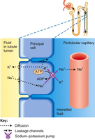
The collecting duct is the final site for water and ion reabsorption, regulated by hormones.
Na+ leakage channels: Allow passive movement of sodium
Principal cells: Respond to aldosterone and ADH
Intercalated cells: Involved in acid-base balance
Hormonal Regulation of Urine Formation
RAA System, ADH, ANP & BNP
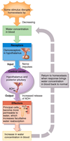
Hormones play a critical role in regulating water and electrolyte balance in the kidneys.
RAA system: Increases Na+ reabsorption and water retention
ADH (antidiuretic hormone): Increases water reabsorption via aquaporin-2 channels
ANP & BNP: Promote natriuresis and diuresis
Urine Concentration
Dilute vs. Concentrated Urine
The kidneys adjust urine concentration based on body needs, using osmotic gradients and permeability changes.
Dilute urine: Produced when water intake is high
Concentrated urine: Produced when water is scarce; involves countercurrent multiplier and urea recycling
Fluid, Electrolyte, and Acid-Base Balance
Fluid Compartments and Balance
Body fluids are distributed between extracellular (ECF) and intracellular (ICF) compartments.
Fluid shift: Movement between compartments
Fluid balance: Input equals output
Electrolyte balance: Maintenance of ion concentrations
Acid-base balance: Regulation of pH
Regulation of Water and Solute Loss
NaCl loss: Can lead to hyponatremia or hypernatremia
Hormonal control: Angiotensin II, aldosterone, ADH, natriuretic peptides
Acid-Base Balance
Acids, Bases, and pH
Acid: Increases [H+] in solution
Base: Decreases [H+] in solution (may form OH- or absorb H+)
Strong vs. weak acids/bases: Strong dissociate completely, weak only partially
Normal pH range: 7.35 – 7.45 (min/max: 6.8/7.7)
Buffer Systems
Buffer systems stabilize pH by absorbing or releasing H+.
Protein buffer system
Phosphate buffer system
Carbonic acid-bicarbonate buffer system:
Acid-Base Disorders
Acidemia: Low blood pH
Alkalemia: High blood pH
Compensation: Complete or partial, respiratory or renal
Respiratory acidosis: Due to hypercapnia (high CO2)
Respiratory alkalosis: Due to hypocapnia (low CO2)
Metabolic acidosis: Causes include lactic acidosis, ketoacidosis, kidney damage, bicarbonate loss
Metabolic alkalosis: Causes include alkaline tide
Summary Table: Urinary System Functions
Function | Mechanism | Regulation |
|---|---|---|
Filtration | Glomerulus | Pressure, GFR |
Reabsorption | PCT, nephron loop, DCT, collecting duct | Hormones (ADH, aldosterone) |
Secretion | DCT, collecting duct | Hormones, transporters |
Acid-base balance | Intercalated cells, buffer systems | Respiratory, renal compensation |
Key Equations
Net Filtration Pressure:
Carbonic Acid-Bicarbonate Buffer:
Additional info:
Expanded explanations of nephron structure and urine formation based on standard textbook content.
Summary table inferred for clarity and completeness.
