 Indietro
Urinary System: Structure, Function, and Clinical Relevance
Guida di studio - Note intelligenti
 Appunti personalizzati basati sui tuoi materiali, ampliati con definizioni chiave, esempi e contesto.
Urinary System Overview
Introduction
The urinary system is essential for maintaining homeostasis by filtering blood, removing waste, and regulating fluid, electrolyte, and acid-base balance. It consists of the kidneys, ureters, urinary bladder, and urethra.
Key Functions: Filtration of blood plasma, elimination of metabolic wastes, regulation of blood volume and pressure, acid-base balance, and secretion of hormones.
Organs: Kidneys, ureters, bladder, urethra.
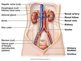
Kidney Anatomy and Location
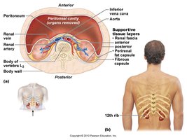
Kidney Structure and Position
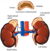
The kidneys are retroperitoneal organs located between T12 and L3 vertebrae, protected by an adipose capsule. Each kidney is about the size of a bar of soap.
Supportive Tissue Layers: Renal fascia, perirenal fat capsule, fibrous capsule.
Location: Posterior abdominal wall, beneath the 12th rib.
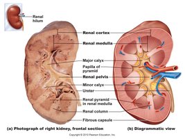
Internal Anatomy of the Kidney
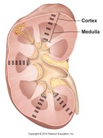
The kidney is divided into the renal cortex and renal medulla. The medulla contains renal pyramids, separated by renal columns. The renal pelvis collects urine from calyces and channels it to the ureter.
Renal Cortex: Outer region, contains nephrons.
Renal Medulla: Inner region, contains pyramids.
Renal Lobe: One pyramid plus overlying cortex.
Major and Minor Calyces: Collect urine from pyramids.
Nephron: Functional Unit of the Kidney
Nephron Structure
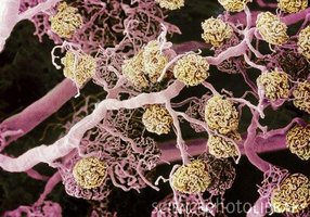
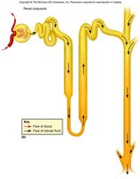
Nephrons are the microscopic functional units responsible for urine formation. Each kidney contains about 1.2 million nephrons.
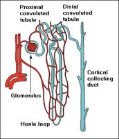
Renal Corpuscle: Includes glomerulus and Bowman’s capsule; site of filtration.
Renal Tubule: Includes proximal convoluted tubule (PCT), nephron loop (Loop of Henle), distal convoluted tubule (DCT), and collecting duct (CD).
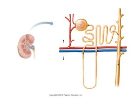
Types of Nephrons
Cortical Nephrons: 85%, short loops, located near kidney surface.
Juxtamedullary Nephrons: 15%, long loops, extend deep into medulla, important for concentrating urine.
Urine Formation
Overview of Urine Formation
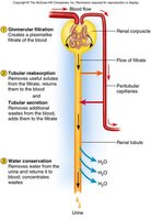
Urine formation involves three main processes: glomerular filtration, tubular reabsorption, and tubular secretion.
Glomerular Filtration: Blood plasma filtered in glomerulus.
Tubular Reabsorption: Useful substances returned to blood.
Tubular Secretion: Additional wastes added to filtrate.
Filtration Membrane
The filtration membrane consists of fenestrated endothelium, basement membrane, and filtration slits formed by podocytes.
Excludes: Blood cells and large molecules (e.g., albumin).
Allows: Water, electrolytes, glucose, amino acids, nitrogenous wastes, vitamins.
Filtration Pressure
Filtration is driven by high blood pressure in the glomerulus, maintained by the wider afferent arteriole compared to the efferent arteriole. 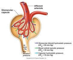
Glomerular Filtration Rate (GFR)
Normal GFR: ≈125 ml/min (male), ≈105 ml/min (female).
99% of filtrate reabsorbed: Only 1–2 L urine excreted/day.
High GFR: Dehydration, electrolyte depletion.
Low GFR: Anuria, waste retention.
Hormonal Regulation of Kidney Function
Renin-Angiotensin-Aldosterone System (RAAS)
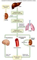
The RAAS regulates blood pressure and fluid balance.
Renin: Released by kidneys in response to low BP.
Angiotensin II: Vasoconstriction, stimulates aldosterone release.
Aldosterone: Promotes Na+ and water reabsorption, increases BP.
Antidiuretic Hormone (ADH)
Released by: Posterior pituitary in response to dehydration.
Effect: Increases water reabsorption in collecting duct, reduces urine volume.
Tubular Reabsorption and Secretion
Reabsorption in Proximal Convoluted Tubule (PCT)
The PCT returns water, glucose, amino acids, and vitamins to the blood.
Solvent Drag: Water absorption by osmosis carries solutes.
Peritubular Capillaries: Favor reabsorption due to high osmolarity and low BP.
Secretion into Tubules
Tubular secretion removes wastes (urea, uric acid, ammonia, drugs) and regulates acid-base balance by secreting H+ and bicarbonate ions. 
Nephron Loop (Loop of Henle)
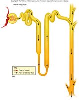
Descending Limb: Water reabsorbed, no electrolytes.
Ascending Limb: Electrolytes reabsorbed (Na+, Cl-, K+), no water.
Salinity Gradient: Allows collecting duct to concentrate urine.
DCT and Collecting Duct
Function: Regulated by hormones (aldosterone, ADH), controls salt/water and acid/base balance.
Medullary CD: More permeable to water, concentrates urine.
Urine Composition and Properties
Normal Urine
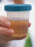
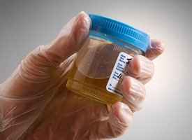
Appearance: Odorless to deep amber, yellow due to urochrome.
Specific Gravity: 1.005–1.030.
Osmolarity: 50–1200 mOsm/L.
pH: 4.5–8.2, usually 6.0.
Chemical Composition: 95% water, 5% solutes (urea, NaCl, KCl, creatinine, uric acid).
Nitrogenous Wastes
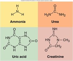
Urea: From amino acid metabolism.
Uric Acid: From nucleic acid catabolism.
Creatinine: From creatine phosphate catabolism.
Urine Volume
Normal: 1–2 L/day.
Polyuria: >2 L/day.
Oliguria: <500 mL/day.
Anuria: 0–100 mL/day.
Diabetes and Diuretics
Diabetes Mellitus: Chronic polyuria, hyperglycemia, glycosuria.
Diabetes Insipidus: ADH hyposecretion, dilute urine.
Diuretics: Increase urine output, decrease blood volume; used for hypertension and heart failure.


Ureters, Bladder, and Urethra
Ureters
Ureters are muscular tubes (about 25 cm long) that transport urine from the renal pelvis to the bladder via peristalsis.
Bladder
The bladder is a distensible organ with a capacity of 500–800 mL. The trigone is a triangular area formed by the openings of the ureters and urethra, susceptible to infection.
Urethra
Female: 3–4 cm, more prone to infections.
Male: 18 cm, internal and external sphincters control flow.
Micturition (Voiding Urine)
Process of Micturition
Stretch receptors in bladder signal the micturition center when bladder contains about 200 mL urine.
Results in contraction of bladder and relaxation of internal sphincter.
Voluntary control via external sphincter.
Urinalysis
Normal and Abnormal Constituents
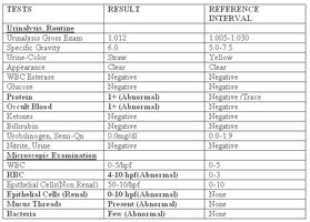
Normal: Urea, creatinine, uric acid, urobilinogen.
Abnormal: Albumin (albuminuria), glucose (glucosuria), blood (hematuria), pus (pyuria), ketones (ketonuria), bilirubin (bilirubinuria), kidney stones, microbes.
Test | Normal Range | Abnormal Finding |
|---|---|---|
Protein | None | Albuminuria |
Glucose | None | Glucosuria |
Blood | None | Hematuria |
Pus | None | Pyuria |
Ketones | None | Ketonuria |
Bilirubin | None | Bilirubinuria |
Microbes | None | UTI |
Kidney Pathology
Common Disorders
Trauma, Infection, Exercise, Hypoxia: Can cause proteinuria or hematuria.
Polycystic Kidney Disease: Genetic, causes cysts, leads to chronic infection and kidney failure.
Kidney Stones: Renal calculi, composed of calcium, phosphate, uric acid, protein; cause pain and obstruction.
Renal Insufficiency: Inability to maintain homeostasis due to nephron destruction.
Renal Cell Carcinoma: Cancer of kidney tubules, symptoms include pain, abnormal urine color, weight loss.
Hemodialysis
Hemodialysis is a treatment for renal failure where blood is filtered through a semipermeable membrane to remove wastes and restore electrolyte balance.
Frequency: Typically 3 sessions/week, 4–8 hours/session.
Kidney Transplant: Most commonly transplanted organ.
Summary Table: Urinary System Functions and Disorders
Function | Disorder | Clinical Significance |
|---|---|---|
Filtration | Glomerulonephritis | Proteinuria, hematuria |
Reabsorption | Diabetes mellitus | Glycosuria, polyuria |
Secretion | Renal insufficiency | Waste retention |
Concentration | Diabetes insipidus | Dilute urine |
Hormonal regulation | Hypertension | Renin/aldosterone imbalance |
Key Equations
Glomerular Filtration Rate (GFR)
Equation:
Net Filtration Pressure (NFP)
Equation:
Conclusion
The urinary system is vital for waste removal, fluid and electrolyte balance, and blood pressure regulation. Understanding its anatomy, physiology, and common pathologies is essential for clinical practice and exam preparation. Review the diagrams and tables for a comprehensive understanding of kidney function and urinary disorders.
