 Indietro
Veins: Structure, Function, and Clinical Significance
Guida di studio - Note intelligenti
 Appunti personalizzati basati sui tuoi materiali, ampliati con definizioni chiave, esempi e contesto.
Veins: Structure and Function
Overview of Veins
Veins are blood vessels responsible for returning blood from the capillaries back to the heart. Compared to arteries, veins have larger diameters, thinner walls, and operate under lower blood pressure. Their structural adaptations facilitate their function in the circulatory system.
Function: Collect blood from capillaries and return it to the heart.
Structural Features: Larger lumens, thinner walls, and lower pressure than arteries.
Clinical Relevance: Disorders such as varicose veins and deep vein thrombosis are associated with venous dysfunction.
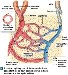
Comparison of Arteries and Veins
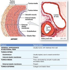
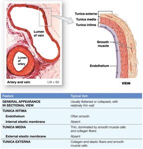
Arteries and veins differ in their structural layers and physical characteristics. Understanding these differences is essential for recognizing their respective roles in circulation.
Arteries: Thick tunica media, round appearance, high pressure.
Veins: Thin tunica media, flattened or collapsed appearance, low pressure.
Key Layers: Both have tunica intima, tunica media, and tunica externa, but the thickness and composition vary.
Feature | Typical Artery | Typical Vein |
|---|---|---|
General Appearance | Round, thick wall | Flattened, thin wall |
Tunica Intima | Ripped (constricted) | Smooth |
Tunica Media | Thick, smooth muscle | Thin, smooth muscle |
Tunica Externa | Collagen, elastic fibers | Collagen, elastic fibers |
Types of Veins
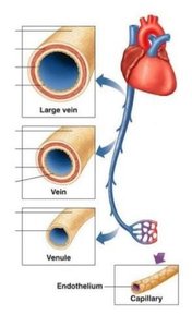
Veins are classified based on their size and function. Each type plays a distinct role in the venous return system.
Venules: Small veins collecting blood from capillaries.
Medium-sized veins: Thin tunica media, few smooth muscle cells, tunica externa with elastic fibers.
Large veins: All three tunica layers, thick tunica externa, thin tunica media; final step in blood return to the heart.
Venous Valves
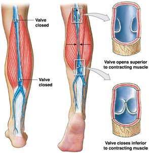
Venous valves are folds of the tunica intima that prevent backflow of blood. Their function is crucial in maintaining unidirectional blood flow toward the heart, especially in the limbs.
Structure: Folds of tunica intima.
Function: Prevent backward flow; compression pushes blood toward the heart.
Clinical Disorders: Weakening of vein walls near valves can lead to varicose veins or hemorrhoids.
Anatomical Location of Veins
Systemic Circuit and Vein Distribution
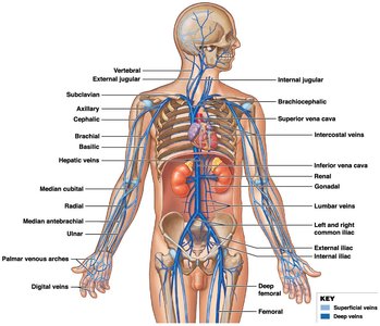
Veins in the systemic circuit run alongside arteries, but their branching patterns are more variable. In the neck and limbs, there are both deep and superficial veins.
Deep veins: Accompany arteries and share their names.
Superficial veins: Located closer to the surface, often used clinically for blood sampling.
Veins of the Upper Limbs
The upper limb contains both deep and superficial veins. Superficial veins are commonly used for venepuncture and intravenous access.
Hand: Superficial palmar arch.
Forearm: Cephalic vein, Basilic vein, Median cubital vein (site for venepuncture).
Arm: Cephalic vein, Basilic vein.
Veins of the Lower Limbs
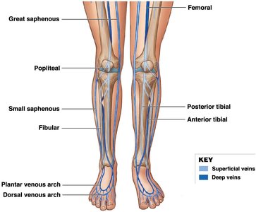
Lower limb veins are also grouped into deep and superficial categories. The great and small saphenous veins are important superficial veins, while deep veins accompany major arteries.
Foot: Dorsal plantar arch.
Leg: Great and small saphenous veins.
Thigh: Great saphenous vein.
Clinical Disorders of the Venous System
Varicose Veins
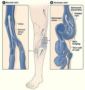
Varicose veins are dilated segments of veins associated with valvular incompetence. They occur when valves fail, allowing blood to flow from deep to superficial veins, causing bulging and discomfort.
Cause: Incompetent valves permit abnormal blood flow.
Location: Occur in superficial veins.
Symptoms: Dilated veins, skin bulging, discomfort.
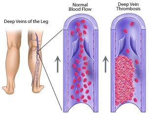
Deep Vein Thrombosis (DVT)
Deep vein thrombosis is the formation of a blood clot within the deep veins of the lower limbs. It can cause pain, swelling, and tenderness, and may lead to pulmonary embolism if the clot travels to the lungs.
Location: Deep veins beneath the fascia of the lower limb.
Complications: Pulmonary embolism, blockage of blood return to the heart.
Symptoms: Pain, swelling, tenderness.
Summary Table: Types and Locations of Veins
Type | Function | Location |
|---|---|---|
Postcapillary Venules | Collect blood from capillaries | Smallest veins, near capillary beds |
Venules | Collect blood from postcapillary venules | Small veins throughout body |
Veins | Return blood to heart | Throughout body, larger vessels |
Superficial Veins | Drain blood from surface tissues | Near skin, limbs |
Deep Veins | Drain blood from deeper tissues | Accompany arteries, limbs |
Key Points to Remember
Veins carry deoxygenated blood from the body to the heart.
Three main types of veins: postcapillary venules, venules, veins.
Two anatomical types: superficial veins and deep veins.
Varicose veins occur in superficial veins; deep vein thrombosis occurs in deep veins.
Additional info: The venous system is essential for maintaining circulatory homeostasis and is a frequent site of clinical intervention due to its accessibility and susceptibility to disease.
