 Indietro
Amino Acid Chemistry, Protein Structure, and Protein Methods
Guida di studio - Note intelligenti
 Appunti personalizzati basati sui tuoi materiali, ampliati con definizioni chiave, esempi e contesto.
Amino Acids: Structure, Properties, and Behavior in Solution
Standard Amino Acids and Their Properties
The 20 standard amino acids are the building blocks of proteins. Each amino acid contains a central carbon (α-carbon) bonded to an amino group, a carboxyl group, a hydrogen atom, and a unique side chain (R group) that determines its properties.
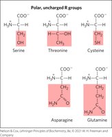
Amino acids differ in their side chains, which can be nonpolar, polar, acidic, or basic.
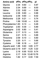
The pKa values of the amino and carboxyl groups, as well as any ionizable side chains, determine the ionization state of the amino acid at different pH values.
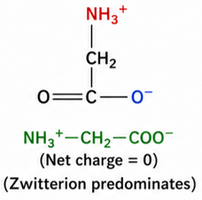
The isoelectric point (pI) is the pH at which the amino acid has no net charge and exists predominantly as a zwitterion.
Ionization and Buffering of Amino Acids
Amino acids can act as buffers due to their ionizable groups. The buffering capacity is greatest near the pKa values of these groups, where both protonated and deprotonated forms are present in significant amounts.
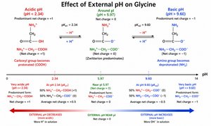
At low pH, amino acids are fully protonated and carry a positive charge.
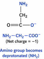
At high pH, they are fully deprotonated and carry a negative charge.
At the pI, the amino acid is a zwitterion with no net charge.
Glycine as an Example
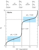
Glycine, the simplest amino acid, has a carboxyl group (pKa1 = 2.34) and an amino group (pKa2 = 9.60). Its pI is calculated as:
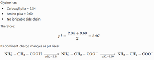
At different pH values, glycine exists in different ionic forms:
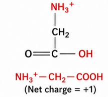
At pH < 2.34: Fully protonated (net charge +1)
At pH ≈ 5.97: Zwitterion (net charge 0)
At pH > 9.60: Fully deprotonated (net charge -1)
Buffering Regions and Titration Curves
The titration curve of glycine shows two buffer regions near its pKa values. The flat regions of the curve indicate effective buffering, while the steep region at the pI indicates poor buffering capacity.
Buffering is most effective when pH ≈ pKa.
The pI is not a buffer region; the amino acid does not resist pH changes well at this point.
Protein Structure: Levels and Stabilizing Interactions
Primary, Secondary, Tertiary, and Quaternary Structure
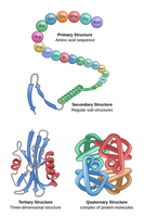
Proteins have four levels of structure, each contributing to their function and stability:
Primary structure: The linear sequence of amino acids in a polypeptide chain, held together by peptide bonds.
Secondary structure: Local folding patterns such as α-helices and β-sheets, stabilized by hydrogen bonds between backbone atoms.
Tertiary structure: The overall three-dimensional shape of a single polypeptide, stabilized by various noncovalent interactions and sometimes disulfide bonds.
Quaternary structure: The assembly of multiple polypeptide subunits into a functional protein complex.
Chemical Bonds and Molecular Interactions in Proteins
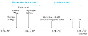
Protein structure is stabilized by both covalent and noncovalent interactions:
Covalent bonds: Peptide bonds (linking amino acids) and disulfide bonds (between cysteine residues).
Noncovalent interactions: Hydrogen bonds, ionic (electrostatic) interactions, hydrophobic interactions, and van der Waals forces.
Peptide Bonds and Protein Size
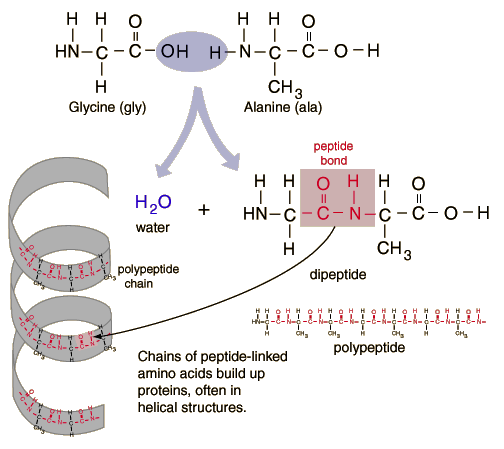
Peptide bonds are covalent bonds formed by condensation reactions between the carboxyl group of one amino acid and the amino group of another. Proteins can range from short peptides to large polypeptides with thousands of residues.
Average molecular weight of an amino acid residue in a protein: 110 Da (after accounting for water loss during peptide bond formation).
Estimating number of residues: Divide protein molecular weight by 110.
Disulfide Bonds
Disulfide bonds are covalent links formed between the sulfhydryl groups of two cysteine residues, resulting in a cystine. These bonds stabilize protein structure, especially in extracellular proteins.
Disulfide bonds are common in secreted proteins, antibodies, and enzymes exposed to harsh environments.
They form in the endoplasmic reticulum (ER) and are rare in cytosolic proteins due to the reducing environment.
Noncovalent Interactions: Ionic, Hydrogen, and Hydrophobic Bonds
Noncovalent interactions are essential for protein folding, stability, and function:
Ionic bonds (salt bridges): Attraction between oppositely charged side chains (e.g., lysine and aspartate).
Hydrogen bonds: Form between backbone atoms in secondary structures and between side chains or with water.
Hydrophobic interactions: Nonpolar side chains cluster away from water, driving protein folding and forming hydrophobic cores.
Protein Purification and Analysis Methods
Protein Separation Techniques
Proteins can be separated and purified based on their physical and chemical properties using chromatography:
Ion-exchange chromatography: Separates proteins by charge.
Size-exclusion (gel filtration) chromatography: Separates proteins by size; large proteins elute first.
Affinity chromatography: Separates proteins by specific binding to a ligand.
Protein Analysis Techniques
After purification, proteins are analyzed for purity, size, and identity:
SDS-PAGE (polyacrylamide gel electrophoresis): Separates proteins by molecular weight.
Western blotting: Detects specific proteins and post-translational modifications using antibodies.
Activity assays: Measure the functional activity of enzymes or proteins.
Summary
Amino acid charge and buffering depend on pH, pKa, and pI values.
Proteins have hierarchical structures stabilized by covalent and noncovalent interactions.
Disulfide bonds and hydrophobic interactions are crucial for protein stability and folding.
Proteins can be separated and analyzed using a variety of biochemical techniques.
