 Indietro
Protein Purification and Analytical Techniques in Biochemistry
Guida di studio - Note intelligenti
 Appunti personalizzati basati sui tuoi materiali, ampliati con definizioni chiave, esempi e contesto.
Protein Analysis and Purification
Overview of Protein Analysis
Understanding protein function and structure requires isolating proteins from complex biological mixtures. This process involves both cellular (in vivo, in cellulo) and isolated-system (in vitro) studies. A complete understanding of a biomolecule's function is achieved by combining these approaches.
In vitro: Study of isolated biomolecules in a test tube.
In vivo: Study within a whole organism.
In cellulo: Study within isolated cells.
Protein purification is essential for functional characterization and must preserve protein activity. The process typically starts with tissue homogenization, followed by separation techniques based on physical and chemical properties.
Chromatography in Organic and Biochemistry
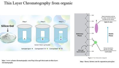
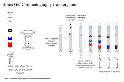
Chromatography is a fundamental technique for separating molecules based on their interactions with stationary and mobile phases. In organic chemistry, silica gel is used as the stationary phase, and separation is based on polarity. In biochemistry, proteins are separated using buffer systems and specialized stationary phases.
Thin Layer Chromatography (TLC): Used for monitoring reaction progress; separation based on polarity.
Silica Gel Chromatography: Used for purifying compounds; less polar molecules move further.
Protein Purification Workflow
Tissue Homogenization and Differential Centrifugation
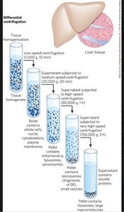
Protein purification begins with tissue homogenization in a buffer, often followed by differential centrifugation to separate cellular components based on size and density. The location of the protein (cytosol, membrane, organelle) determines the centrifugation protocol.
Low-speed centrifugation: Pellets large components (cells, nuclei).
Medium-speed centrifugation: Pellets mitochondria, lysosomes.
High-speed centrifugation: Pellets small vesicles.
Ultracentrifugation: Isolates soluble proteins.
Ammonium Sulfate Precipitation (Salting Out)
Proteins differ in solubility at various ionic strengths. Ammonium sulfate precipitation exploits this by causing proteins to precipitate at high salt concentrations, a process known as "salting out." This is a cost-effective early purification step.
Protein Chromatography Techniques
Ion Exchange Chromatography
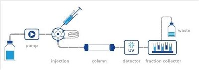
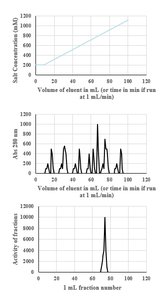
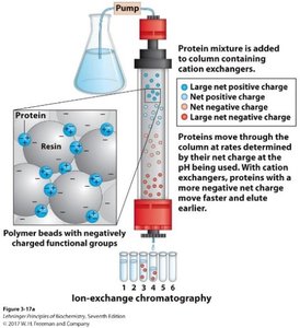
Ion exchange chromatography separates proteins based on net charge. The stationary phase can be anionic (cation exchange) or cationic (anion exchange). The protein's isoelectric point (pI) determines its interaction with the column.
Cation exchange: Anionic stationary phase; cationic proteins bind tightly.
Anion exchange: Cationic stationary phase; anionic proteins bind tightly.
Elution: Achieved by salt gradient or pH change; salt competes with protein for binding.
Specific Activity and Protein Quantification
Specific activity is defined as enzyme activity per unit of protein mass. It increases as purification progresses, even if total activity decreases, because contaminating proteins are removed. The Bradford assay is commonly used to quantify total protein.
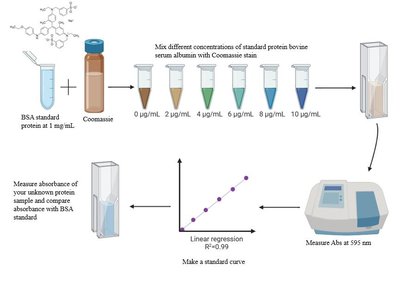
Bradford assay: Uses Coomassie dye; absorbance measured at 595 nm.
Standard curve: Created using known concentrations of bovine serum albumin (BSA).
Size Exclusion Chromatography (SEC)
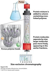
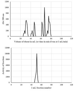
SEC separates proteins based on size. The stationary phase contains porous beads; small proteins enter the pores and elute later, while large proteins elute earlier. SEC preserves quaternary structure and is used to estimate native molecular weight.
Analytical use: Compare elution volume to standards to infer oligomeric state.
Affinity Chromatography
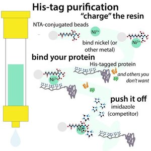
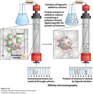
Affinity chromatography exploits specific binding interactions between a protein and a ligand. Recombinant proteins can be tagged (e.g., His-tag) for immobilized metal affinity chromatography (IMAC). Alternatively, a ligand (such as NADH) can be coupled to a resin to capture proteins with specific binding properties.
IMAC: His-tagged proteins bind to Ni²⁺ or Co²⁺ resin; eluted with imidazole.
Ligand-based affinity: Ligand coupled to resin captures target protein; eluted with free ligand.
Protein Analytical Methods
SDS-PAGE (Sodium Dodecyl Sulfate Polyacrylamide Gel Electrophoresis)
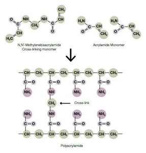
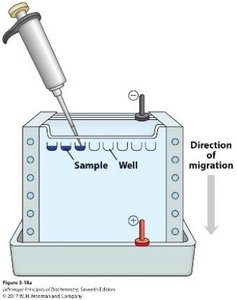
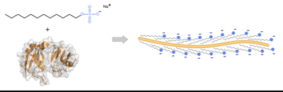
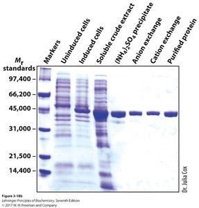
SDS-PAGE separates proteins by size. SDS denatures proteins and imparts a uniform negative charge, allowing separation based solely on molecular weight. The gel is formed from polyacrylamide, and proteins migrate toward the anode.
SDS: Denatures proteins, coats them with negative charge.
Reducing agent: β-mercaptoethanol reduces disulfide bonds.
Analysis: Purity assessed by number of bands; quaternary structure inferred by comparing SEC and SDS-PAGE results.
Isoelectric Focusing
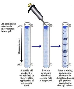
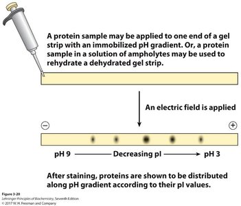
Isoelectric focusing separates proteins based on their isoelectric point (pI). A pH gradient is established in the gel, and proteins migrate until their net charge is zero, stopping at their pI.
Ampholytic solution: Forms pH gradient in gel.
Analysis: Multiple proteins with different pI can be separated.
2D-Gel Electrophoresis
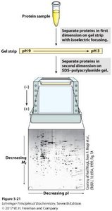
2D-gel electrophoresis combines isoelectric focusing and SDS-PAGE, separating proteins first by pI and then by size. This technique provides high-resolution analysis and is often used before protein sequencing.
Protein Sequencing Techniques
Edman Degradation
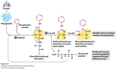
Edman degradation sequentially removes N-terminal amino acids, which are identified by HPLC or LC-MS. Partial sequence data can be matched to genomic databases to identify the full-length protein.
Protease digestion: Trypsin, chymotrypsin, and CNBr generate fragments for sequencing.
Sequence reconstruction: Overlapping fragments are used to deduce the full sequence.
Mass Spectrometry for Protein Sequencing
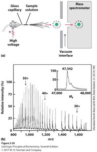
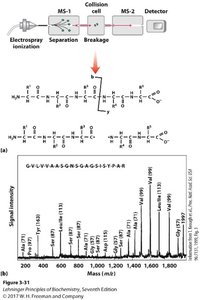
Mass spectrometry (MS) is the modern standard for protein sequencing. Electrospray ionization (ESI) produces multiply charged ions, and tandem MS (MS/MS) fragments peptides to determine sequence.
ESI-MS: Measures mass-to-charge ratio; produces multiple charge states.
LC-MS/MS: Peptides are separated, fragmented, and sequenced based on mass differences.
Summary Table: Protein Purification Steps and Properties
Step | Property Exploited | Technique |
|---|---|---|
Tissue Homogenization | Cell disruption | Blending, buffer |
Differential Centrifugation | Size, density | Centrifugation |
Salting Out | Solubility | Ammonium sulfate precipitation |
Ion Exchange Chromatography | Net charge, pI | Cation/anion exchange |
Size Exclusion Chromatography | Size | SEC |
Affinity Chromatography | Specific binding | IMAC, ligand-based |
SDS-PAGE | Size (denatured) | Electrophoresis |
Isoelectric Focusing | pI | Electrophoresis |
2D-Gel Electrophoresis | pI and size | Combined electrophoresis |
Edman Degradation | N-terminal sequence | Chemical cleavage |
Mass Spectrometry | Mass, sequence | ESI-MS, LC-MS/MS |
Key Equations
Specific Activity:
Fold Purification:
Additional info:
Some context and examples were expanded for clarity and completeness, including clinical applications and workflow integration. Images were included only when directly relevant to the explanation of the adjacent paragraph.
