Indietro
Protein Structure, Folding, and Function: Advanced Study Notes
Guida di studio - Note intelligenti
 Appunti personalizzati basati sui tuoi materiali, ampliati con definizioni chiave, esempi e contesto.
Protein Folding: Principles and Mechanisms
Rules and Observations for Protein Folding
Protein folding is a highly regulated process driven primarily by the hydrophobic effect, which organizes the polypeptide chain into its functional three-dimensional structure.
Hydrophobic Effect: Hydrophobic residues are buried in the protein core, while hydrophilic residues remain on the surface, resulting in globular proteins with two structural layers.
Secondary Structure Segregation: Alpha helices and beta sheets, when present together, are typically found in separate layers due to limited hydrogen bonding between these elements.
Beta Sheet Stability: Beta sheets are most stable with a right-handed twist. Antiparallel beta sheets often contain beta bulges—imperfections where one strand has an extra amino acid compared to its neighbor.
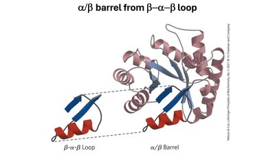
Motif Assembly: Common motifs, such as the beta-alpha-beta loop, can fold into more complex structures like the alpha/beta barrel.
Intrinsically Disordered Proteins (IDPs)
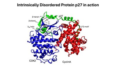
Intrinsically disordered proteins (IDPs) lack a stable tertiary structure under physiological conditions but can contain regions of secondary structure. Despite their flexibility, IDPs perform essential cellular functions.
Structural Features: IDPs are rich in amino acids such as lysine (K), arginine (R), aspartic acid (D), glutamic acid (E), and proline (P).
Functional Roles: They act as spacers, linkers, and scavengers for ions or small molecules, and can interact with multiple targets due to their flexibility.
Classification and Higher-Order Structure of Proteins
Protein Classification
Proteins are classified based on their folding patterns and sequence similarities. The SCOP2 database organizes proteins into families and superfamilies:
Family: Proteins with similar sequences and folding patterns.
Superfamily: Proteins with similar folding patterns but low sequence similarity.
Evolutionary Insight: Some families are conserved across all domains of life, indicating ancient origins, while others are restricted to specific lineages.
Quaternary Structure
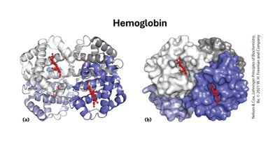
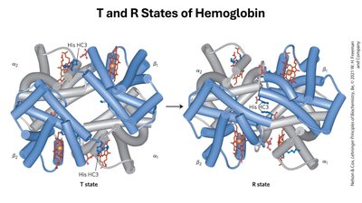
Quaternary structure refers to the assembly of multiple polypeptide chains (subunits) into a functional protein complex. Hemoglobin is a classic example:
Hemoglobin: A tetramer composed of two alpha and two beta subunits (a dimer of alpha/beta protomers), each containing a heme prosthetic group (protoporphyrin and Fe(II)).
Protein Folding and Denaturation
Energetics and Cooperativity
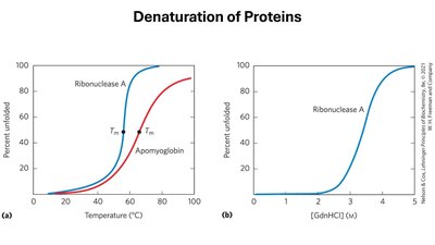
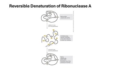
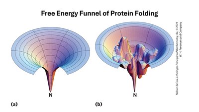
The energy difference between the folded (native) and unfolded (denatured) states of a protein is small, making folding and denaturation highly cooperative processes. Proteostasis refers to the cellular maintenance of functional proteins.
Denaturation: Can be induced by heat, pH extremes, high salt, urea, guanidinium, organic solvents, or detergents. It is a highly cooperative process, often studied by circular dichroism (CD).
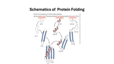
Folding: Also highly cooperative, with tertiary and quaternary structures encoded in the primary sequence.
Protein Function: Ligand Binding and Cooperativity
Ligand Binding Equilibria
Many proteins function by reversibly binding ligands. The binding equilibrium can be described as follows:
Equilibrium:
Association Constant:
Dissociation Constant:
Fraction Bound (Y):
Operational Definition of : When , (half of the protein is ligand-bound).
Cooperativity and the Hill Equation
Proteins with multiple binding sites can exhibit cooperativity, described by the Hill equation:
Hill Equation: , where is the Hill coefficient.
Types of Cooperativity: Positive (), Negative ().
Hill Plot:
Globins: Structure and Function
Types of Globins
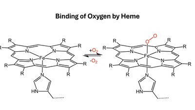
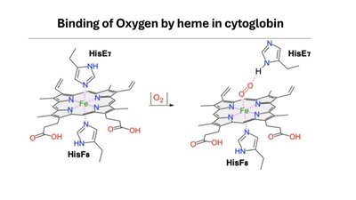
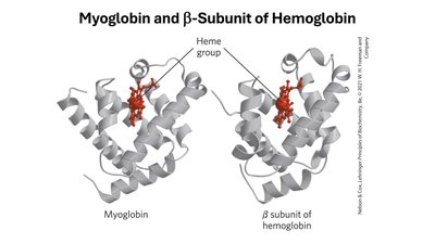
Globins are a superfamily of proteins involved in oxygen transport and storage. In humans, there are four main types:
Myoglobin: Stores oxygen in muscle; single polypeptide with a heme group; slow O2 binding.
Neuroglobin: Found in neural tissues; protects neurons under hypoxia.
Cytoglobin: Found in blood vessel walls; binds O2 and NO, regulating muscle relaxation.
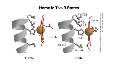
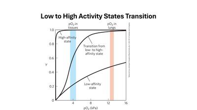
Hemoglobin: Oxygen carrier in blood; tetrameric structure; exhibits cooperative O2 binding.
Protein-Protein Interactions: Immune System and Muscle Contraction
Immune System: Antibodies and Antigens
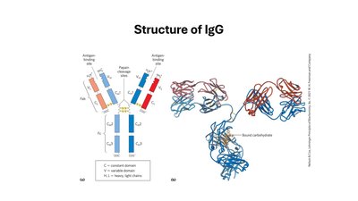
The immune system relies on protein-protein interactions for defense. Antibodies (immunoglobulins) are produced by B cells and recognize specific antigens (foreign molecules).
Antibody Structure: Y-shaped molecules with two heavy and two light chains; each chain has constant and variable domains.
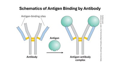
Antigen Binding: Occurs at the variable domains; each antibody has two identical binding sites.
Antigenic Determinant (Epitope): The specific region of the antigen recognized by the antibody.
Haptens: Small molecules that can elicit an immune response only when attached to a larger carrier.
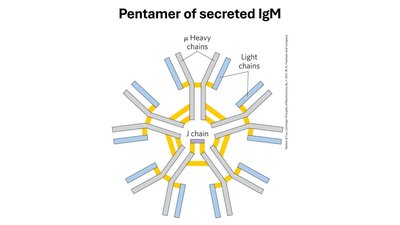
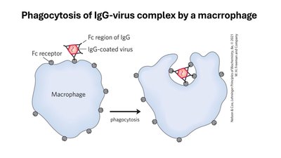
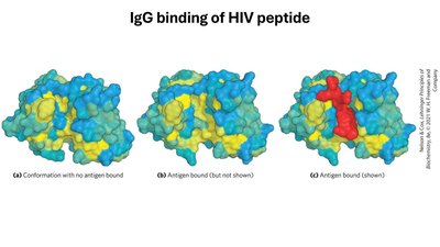
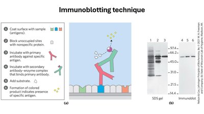
Types of Immunoglobulins
Type | Heavy Chain | Structure | Function |
|---|---|---|---|
IgG | Gamma (γ) | Monomer | Most abundant in plasma; secondary response; activates macrophages |
IgM | Mu (μ) | Pentamer (secreted), Monomer (membrane) | Primary response antibody |
IgA | Alpha (α) | Monomer, Dimer, Trimer | Secreted in fluids (saliva, tears, milk) |
IgD | Delta (δ) | Monomer | Regulates IgM activity |
IgE | Epsilon (ε) | Monomer | Allergic responses; activates mast cells |
Analytical Applications
Polyclonal Antibodies: Produced by multiple B cells; recognize multiple epitopes.
Monoclonal Antibodies: Produced by identical B cells; recognize a single epitope; used in immunoblotting (Western blot).
Muscle Contraction: Myosin and Actin
Muscle contraction is driven by the interaction of myosin and actin, powered by chemical energy (ATP hydrolysis).
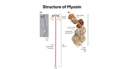
Myosin Structure: Hexamer with three heavy chains and associated light chains; the tail forms a left-handed supercoil, and the heads interact with actin filaments.