Indietro
A Tour of the Cell: Structure, Function, and Organization
Guida di studio - Note intelligenti
 Appunti personalizzati basati sui tuoi materiali, ampliati con definizioni chiave, esempi e contesto.
Chapter 6: A Tour of the Cell
Introduction to Cell Biology
The cell is the fundamental unit of structure and function in all living organisms. Understanding cell structure and function is essential for studying biology at all levels, from molecular to ecological.
Cells are the smallest units of life capable of performing all vital functions.
Organisms may be unicellular or multicellular, but all are composed of cells.
Concept 6.1: Biologists Use Microscopes and Biochemistry to Study Cells
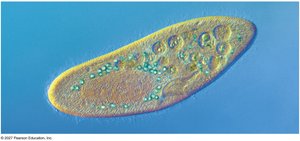
Cells are typically too small to be seen without magnification. Microscopy and biochemical techniques are essential for cell study.
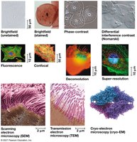
Microscopy allows visualization of cells and their components.
Three key parameters: magnification (image size vs. real size), resolution (clarity), and contrast (brightness differences).
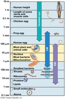
Light microscopes use visible light and glass lenses to magnify specimens.
Electron microscopes provide higher resolution for detailed cell structure.
Cell Fractionation
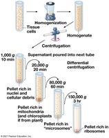
Cell fractionation is a technique used to separate cellular components for study.
Cells are homogenized and then centrifuged to separate organelles by size and density.
This enables scientists to determine the functions of individual organelles.
Concept 6.2: Eukaryotic Cells Have Internal Membranes That Compartmentalize Their Functions
Prokaryotic vs. Eukaryotic Cells
All cells share basic features, but prokaryotic and eukaryotic cells differ in complexity and organization.
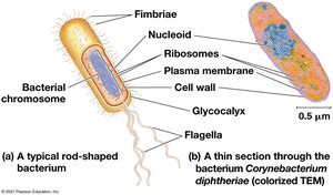
Basic features: plasma membrane, cytosol, chromosomes, ribosomes.
Prokaryotic cells (Bacteria, Archaea): No nucleus, DNA in nucleoid, no membrane-bound organelles.
Eukaryotic cells (Protists, fungi, animals, plants): DNA in nucleus, membrane-bound organelles, larger size.
Cell Size and Surface Area-to-Volume Ratio
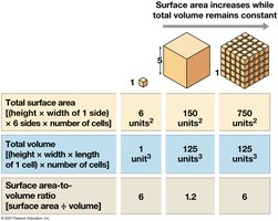
Cell size is limited by metabolic requirements and the surface area-to-volume ratio.
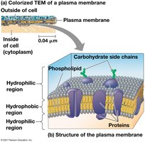
The plasma membrane controls passage of materials.
As cell size increases, volume grows faster than surface area, affecting efficiency.
A Panoramic View of the Eukaryotic Cell
Eukaryotic cells contain internal membranes and organelles that compartmentalize functions.
Membranes create distinct environments for specialized processes.
Biological membranes are primarily composed of phospholipids.
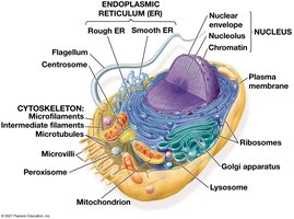
Concept 6.3: The Eukaryotic Cell’s Genetic Instructions Are Housed in the Nucleus and Carried Out by the Ribosomes
The Nucleus: Information Central
The nucleus stores genetic information and coordinates cellular activities.
DNA is organized into chromosomes, associated with proteins as chromatin.
The nucleolus synthesizes ribosomal RNA (rRNA).
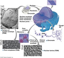
Ribosomes: Protein Factories
Ribosomes are responsible for protein synthesis.
Composed of rRNA and protein.
Located in cytosol (free ribosomes) or attached to endoplasmic reticulum/nuclear envelope (bound ribosomes).
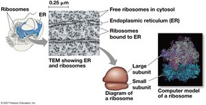
Concept 6.4: The Endomembrane System Regulates Protein Traffic and Performs Metabolic Functions
Components of the Endomembrane System
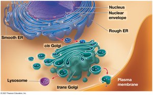
The endomembrane system includes several interconnected organelles that manage protein and lipid synthesis, transport, and degradation.
Nuclear envelope, endoplasmic reticulum (ER), Golgi apparatus, lysosomes, vacuoles, plasma membrane.
Components are connected directly or via vesicles.
The Endoplasmic Reticulum (ER)
The ER is a biosynthetic factory with two regions: smooth and rough.
Smooth ER: Synthesizes lipids, detoxifies drugs, stores calcium ions.
Rough ER: Studded with ribosomes, synthesizes glycoproteins, distributes transport vesicles, produces membranes.
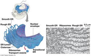
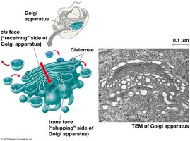
The Golgi Apparatus: Shipping and Receiving Center
The Golgi apparatus modifies, sorts, and packages proteins and lipids for transport.
Consists of stacked cisternae.
Receives products from ER, processes them, and sends them to their destinations.
Lysosomes: Digestive Compartments
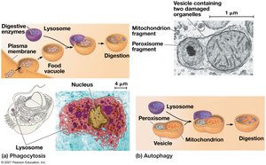
Lysosomes contain hydrolytic enzymes for digestion and recycling.
Digest macromolecules and damaged organelles.
Formed by rough ER and processed by Golgi apparatus.
Engage in phagocytosis and autophagy.
Vacuoles: Diverse Maintenance Compartments
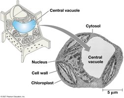
Vacuoles are large vesicles with varied functions in different cells.
Food vacuoles (phagocytosis), contractile vacuoles (water regulation), central vacuole (storage and growth in plants).
Concept 6.5: Mitochondria and Chloroplasts Change Energy from One Form to Another
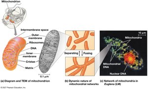
Mitochondria: Chemical Energy Conversion
Mitochondria are the sites of cellular respiration, converting oxygen and nutrients into ATP.
Double membrane structure, inner membrane forms cristae.
Matrix contains enzymes for metabolic reactions.
Chloroplasts: Capture of Light Energy
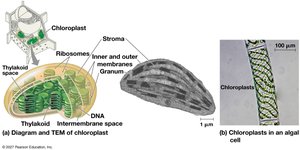
Chloroplasts are the sites of photosynthesis in plants and algae.
Contain chlorophyll, thylakoids (stacked as grana), and stroma.
Part of the plastid family of organelles.
Peroxisomes: Oxidation
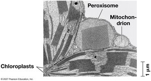
Peroxisomes are specialized organelles for oxidative reactions.
Break down fatty acids, detoxify harmful substances, convert fatty acids to sugars in plant seeds.
Endosymbiont Theory
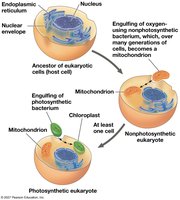
The endosymbiont theory explains the evolutionary origin of mitochondria and chloroplasts.
Suggests eukaryotic cells arose from engulfing prokaryotic cells.
Evidence: double membranes, own DNA, ribosomes, independent reproduction.
Concept 6.6: The Cytoskeleton Is a Network of Fibers That Organizes Structures and Activities in the Cell
Cytoskeleton Structure and Function
The cytoskeleton provides structural support, organization, and motility for cells.
Three types: microtubules, microfilaments, intermediate filaments.
Anchors organelles and facilitates movement.
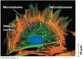
Roles of the Cytoskeleton: Support and Motility
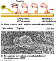
The cytoskeleton interacts with motor proteins to move organelles and vesicles.
Motor proteins "walk" along cytoskeletal tracks using ATP.
Microfilaments and Cellular Motility
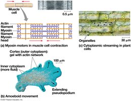
Microfilaments, composed of actin, are involved in cell movement and shape changes.
Work with myosin for muscle contraction and cytoplasmic streaming.
Enable amoeboid movement via pseudopodia.
Intermediate Filaments
Intermediate filaments provide structural stability and anchor organelles.
More permanent than microtubules or microfilaments.
Concept 6.7: Extracellular Components and Connections Between Cells Help Coordinate Cellular Activities
Cell Walls of Plants
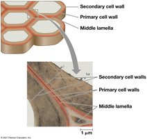
Plant cells are distinguished by their rigid cell walls, which provide protection and structural support.
Made of cellulose, polysaccharides, and proteins.
Multiple layers: primary cell wall, middle lamella, secondary cell wall.
Extracellular Matrix (ECM) of Animal Cells
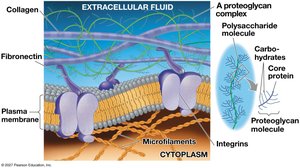
Animal cells are surrounded by an extracellular matrix (ECM) that provides structural and biochemical support.
Composed of glycoproteins (collagen, proteoglycans, fibronectin).
Integrins connect ECM to the cytoskeleton.
Cell Junctions
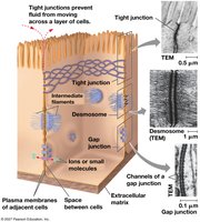
Cells in tissues are connected by specialized junctions that facilitate communication and adhesion.
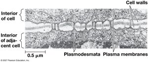
Plasmodesmata (plants): Channels for transport between cells.
Tight junctions (animals): Prevent fluid leakage.
Desmosomes: Anchor cells together.
Gap junctions: Allow passage of ions and molecules.
Concept 6.8: A Cell Is Greater Than the Sum of Its Parts
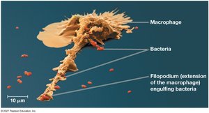
Cellular functions depend on the coordinated activity of all components. For example, immune cells use cytoskeleton, lysosomes, and plasma membrane to engulf and destroy bacteria.