 Indietro
A Tour of the Cell: Structure, Function, and Organization
Guida di studio - Note intelligenti
 Appunti personalizzati basati sui tuoi materiali, ampliati con definizioni chiave, esempi e contesto.
A Tour of the Cell
Microscopy and Studying Cells
Cells are the fundamental units of life, and their study requires specialized tools such as microscopes due to their small size. Microscopy allows scientists to observe cell structure, function, and diversity.
Cell size and organelles correlate with function.
All cells contain nucleic acids, proteins, carbohydrates, and a plasma membrane.
Cells are dynamic, with interacting parts and constantly moving molecules.
Example: A single-celled organism such as Paramecium demonstrates internal complexity and dynamic processes.
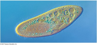
Most cells are microscopic and require magnification to be seen. Bacteria are the smallest cells, while plant and animal cells are about ten times larger.
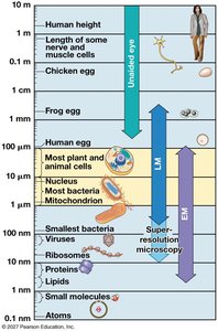
Key Point: Cell size and internal organization are directly tied to function.
Prokaryotic and Eukaryotic Cells
Cells are classified into two broad groups based on morphology: prokaryotes and eukaryotes. Phylogenetically, life is divided into three domains: Bacteria, Archaea (both prokaryotic), and Eukarya (eukaryotic).
Prokaryotes: Lack a membrane-bound nucleus; DNA is found in the nucleoid region.
Eukaryotes: Have a nucleus surrounded by a double membrane and possess membrane-bound organelles.
Example: The structure of a typical prokaryotic cell includes the nucleoid, ribosomes, plasma membrane, cell wall, and sometimes flagella and fimbriae.
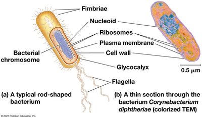
Comparison Table:
Feature | Prokaryotes | Eukaryotes |
|---|---|---|
Location of DNA | Nucleoid (not membrane bound); plasmids common | Inside nucleus (membrane bound); plasmids rare |
Internal Membranes/Organelles | Limited; extensive only in photosynthetic species | Many types and numbers of organelles |
Cytoskeleton | Limited | Extensive |
Overall Size | Usually small | Larger |
Metabolic requirements set upper limits on cell size, and compartmentalization in eukaryotes separates incompatible reactions and increases efficiency.
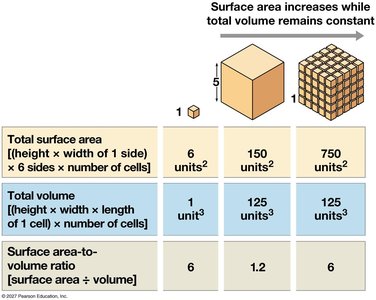
Eukaryotic Cell Structure and Compartmentalization
Eukaryotic cells are partitioned into functional compartments, each specialized for particular life processes.
Manufacturing: Nucleus, ribosomes, endoplasmic reticulum, Golgi apparatus
Breakdown: Lysosomes, vacuoles, peroxisomes
Energy processing: Mitochondria (animal cells), chloroplasts (plant cells)
Structural support, movement, communication: Cytoskeleton, plasma membrane, cell wall
Example: Generalized animal cell showing major organelles and structures.
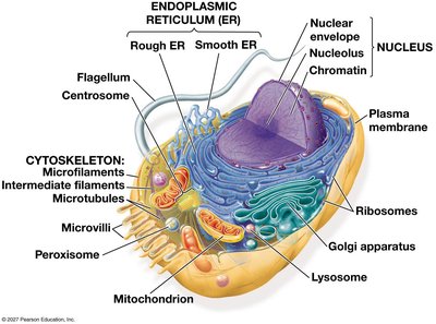
Compartmentalization offers two primary advantages: separation of incompatible chemical reactions and increased efficiency of chemical reactions.
The Nucleus and Ribosomes
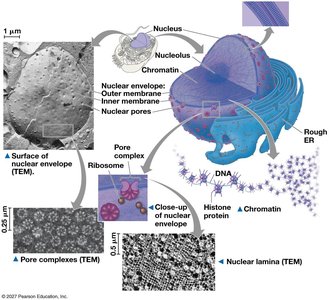
The nucleus is the information center of the cell, housing most of the cell's genes. It is surrounded by a double membrane (nuclear envelope) with nuclear pores for molecular transport.
Nuclear lamina: Maintains nuclear shape
Nuclear matrix: Protein framework inside nucleus
Nucleolus: Site of ribosomal RNA (rRNA) synthesis
Chromatin: DNA associated with proteins
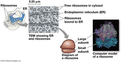
Ribosomes are non-membranous complexes responsible for protein synthesis. They consist of large and small subunits, both containing RNA and protein. Ribosomes can be free in the cytosol or bound to the rough ER.

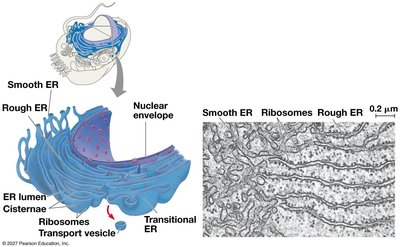
The Endomembrane System
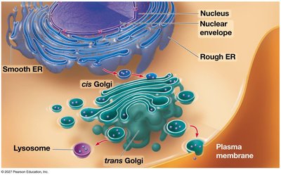
The endomembrane system is a coordinated network of organelles involved in synthesis, processing, and transport of proteins and lipids.
Rough ER: Studded with ribosomes; synthesizes proteins for secretion, membrane, or organelles.
Smooth ER: Lacks ribosomes; synthesizes lipids, detoxifies, stores Ca2+.
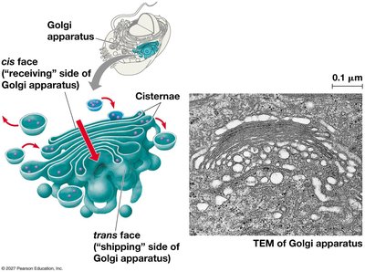
Golgi apparatus: Stacked cisternae; processes, sorts, and ships proteins from rough ER.
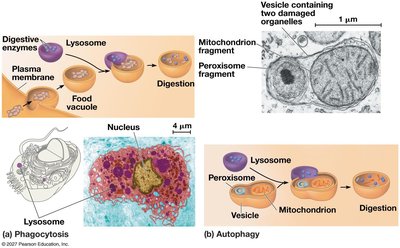
Lysosomes: Single-membrane-bound; contain digestive enzymes for waste processing (animal cells).
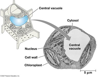
Vacuoles: Large, membrane-bound; digestion, storage, water balance (plants, fungi).
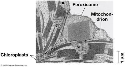
Peroxisomes: Globular, single membrane; center of oxidation reactions.
Mitochondria and Chloroplasts
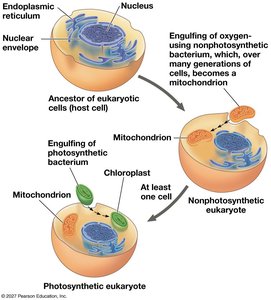
Mitochondria and chloroplasts are energy-processing organelles believed to have originated via endosymbiosis.
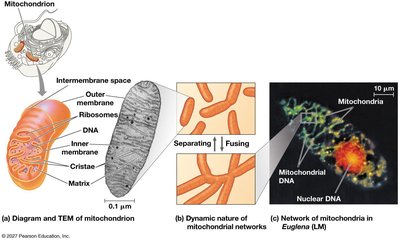
Mitochondria: Double membrane, own DNA/ribosomes, replicate by fission; core function is ATP production.
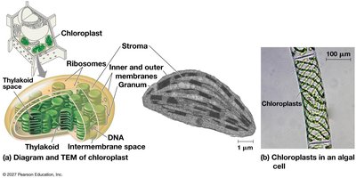
Chloroplasts: Double membrane, thylakoids stacked into grana, own DNA/ribosomes; convert light energy to chemical energy via photosynthesis.
The Cytoskeleton
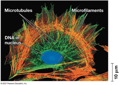
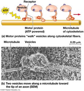
The cytoskeleton is a network of protein fibers that provides structural support, organizes cell contents, and enables movement.
Actin filaments (microfilaments): Support cell shape, drive movement with myosin motor proteins.
Intermediate filaments: Provide structural support (e.g., keratin, nuclear lamins).
Microtubules: Hollow tubes, tracks for vesicle transport, power cilia/flagella.
Extracellular Structures and Cell Junctions
Cells are supported and connected by extracellular structures and junctions.
Cell wall: Provides protection and structural support in plants (cellulose), fungi (chitin), some protists.
Extracellular matrix (ECM): Surrounds animal cells, made of glycoproteins (collagen, proteoglycans, fibronectin), binds cells via integrins.
Cell junctions: Tight junctions seal, desmosomes anchor, gap junctions connect animal cells.
The Cell as an Integrated Whole
Cellular life is an emergent property arising from the cooperation of organelles and structures. Organelles do not work in isolation; their functions are interdependent.
Example: Phagocytosis by a macrophage requires the cytoskeleton, plasma membrane, and lysosomes acting together.
Mitochondria are involved in ATP production, cell signaling, and hormone synthesis.
Organelle Quick Reference
Organelle | Function |
|---|---|
Nucleus | Information storage/transmission; ribosome subunit assembly |
Ribosomes | Protein synthesis |
Rough ER | Protein synthesis and processing |
Smooth ER | Lipid synthesis and processing; detoxification |
Golgi apparatus | Protein, lipid, and carbohydrate processing |
Lysosomes | Digestion and recycling |
Vacuoles | Storage; digestion; water balance |
Peroxisomes | Oxidation of fatty acids and other compounds |
Mitochondria | ATP production |
Chloroplasts | Photosynthesis — production of sugars from light energy |
Cytoskeleton | Structural support; movement of materials and the cell itself |
Plasma membrane | Selective permeability; maintains intracellular environment |
Cell wall | Protection and structural support (plants, fungi, some protists) |
Extracellular matrix | Binds cells in tissues; regulates cellular activity |
Cell junctions | Communication and binding between neighboring cells |
Additional info: The notes above expand on brief points with academic context, definitions, and examples to provide a self-contained study guide for General Biology students.
