 Indietro
A Tour of the Cell: Structure, Function, and Diversity
Guida di studio - Note intelligenti
 Appunti personalizzati basati sui tuoi materiali, ampliati con definizioni chiave, esempi e contesto.
Introduction to the Cell
The Cell as the Fundamental Unit of Life
Cells are the basic structural and functional units of all living organisms. Life emerges at the cellular level, and the similarities among all cells demonstrate the unity of life, while their differences reveal its diversity.
All organisms are made of cells
Cells are the fundamental unit of life
Unity and diversity: Similarities show unity; differences show diversity
Types of Cells
Prokaryotic vs. Eukaryotic Cells
Cells are classified into two main types: prokaryotic and eukaryotic. This classification is based on the presence or absence of membrane-bound organelles.
Prokaryotic cells: Found in Bacteria and Archaea domains; lack membrane-bound organelles and a nucleus
Eukaryotic cells: Found in Eukarya domain (protists, fungi, animals, plants); have membrane-bound organelles and a nucleus
Basic Features of All Cells
Plasma membrane: Boundary of the cell
Cytosol: Semifluid substance inside the cell
Chromosomes: Carry genetic information
Ribosomes: Synthesize proteins
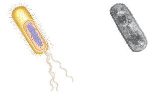
Prokaryotic Cell Structure
No membrane-bound organelles
No nucleus; DNA is in the nucleoid region
Cytoplasm bound by the plasma membrane
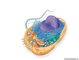
Eukaryotic Cell Structure
Has a nucleus with DNA bound by a nuclear envelope
Contains membrane-bound organelles
Cytoplasm is the region between the plasma membrane and nucleus
Generally larger than prokaryotic cells
Cell Size and Surface Area
Metabolic Requirements and Cell Size
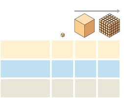
Cell size is limited by metabolic requirements. The plasma membrane is critical for the exchange of oxygen, nutrients, and waste. As a cell increases in size, its volume grows faster than its surface area, affecting its efficiency.
Surface area-to-volume ratio is important for cell function
Cells must maintain sufficient surface area for exchange
Internal Membranes and Organelles
Compartmentalization in Eukaryotic Cells
Internal membranes in eukaryotic cells partition the cell into discrete environments, allowing specialized functions. Biological membranes are composed of a double layer of phospholipids and embedded proteins.
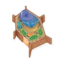
Plant and animal cells share most organelles
Membranes create functional compartments
Cell Diversity
Examples of Eukaryotic Cells
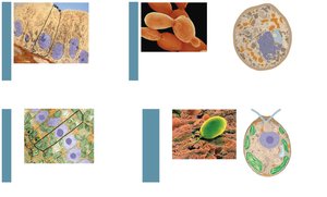
Eukaryotic cells can be animal, fungal, plant, or unicellular protists. Each type has unique features and organelles.
Animal cells: No cell wall, have lysosomes
Fungal cells: Cell wall, vacuole, mitochondria
Plant cells: Cell wall, chloroplasts, central vacuole
Unicellular eukaryotes: Diverse structures
The Nucleus
Structure and Function
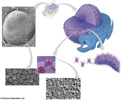
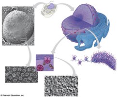
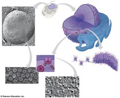
The nucleus is the control center of the cell, containing most of the cell's DNA. It is surrounded by a double-layered nuclear envelope with nuclear pores for transport.
Nuclear envelope: Double membrane with pores
Nuclear lamina: Protein filaments supporting the envelope
Nuclear matrix: Internal framework
Chromosomes: DNA and proteins, condense during division
Nucleolus: Site of rRNA synthesis and ribosome assembly
Ribosomes
Protein Synthesis
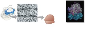
Ribosomes are not organelles; they are complexes of rRNA and protein that carry out protein synthesis. They exist as free ribosomes in the cytosol or bound to the ER or nuclear envelope.
Free ribosomes: Synthesize cytosolic proteins
Bound ribosomes: Synthesize membrane-bound or secretory proteins
The Endomembrane System
Components and Functions
The endomembrane system regulates protein traffic and performs metabolic functions. It includes the nuclear envelope, ER, Golgi apparatus, lysosomes, vesicles, vacuoles, and plasma membrane.
Components are connected or transfer materials via vesicles
Highly diverse in function and structure
Endoplasmic Reticulum (ER)
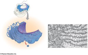
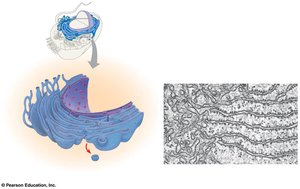
The ER is a network of membranous tubules and sacs (cisternae) continuous with the nuclear envelope. It separates the cytosol from the ER lumen.
Smooth ER: Lacks ribosomes; synthesizes lipids, metabolizes carbohydrates, detoxifies drugs, stores calcium
Rough ER: Studded with ribosomes; produces secretory proteins and membranes
Golgi Apparatus
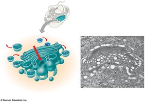
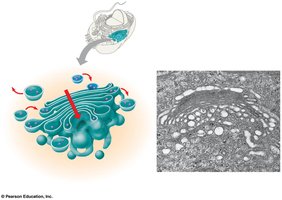
The Golgi apparatus modifies, sorts, and packages products from the ER. It consists of flattened sacs called cisternae, with a cis face (receiving) and trans face (shipping).
Manufactures macromolecules
Modifies glycoproteins
Tags products for final destination
Lysosomes
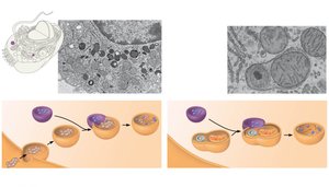
Lysosomes are membranous sacs of hydrolytic enzymes that digest macromolecules. They function in phagocytosis and autophagy, recycling cellular components.
Work best in acidic environments
Digest food vacuoles and damaged organelles
Vacuoles
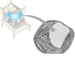
Vacuoles are large vesicles derived from the ER and Golgi. They perform various functions, including storage, water regulation, and enzymatic hydrolysis.
Food vacuoles: Formed by phagocytosis
Contractile vacuoles: Pump excess water
Central vacuole: Stores organic compounds and water in plants
Mitochondria and Chloroplasts
Energy Conversion Organelles
Mitochondria and chloroplasts convert energy from one form to another. Mitochondria are sites of cellular respiration, while chloroplasts are sites of photosynthesis.
Mitochondria: Use oxygen to generate ATP
Chloroplasts: Convert solar energy to chemical energy
Endosymbiont Theory
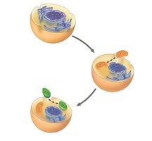
This theory proposes that mitochondria and chloroplasts originated from prokaryotes engulfed by ancestral eukaryotes. They are similar to bacteria in structure and function.
Double membrane
Free ribosomes and circular DNA
Semiautonomous: grow and reproduce independently
Mitochondria Structure
Double membrane: smooth outer, folded inner (cristae)
Intermembrane space and mitochondrial matrix
Contains enzymes, mtDNA, and ribosomes
Chloroplast Structure
Double membrane with intermembrane space
Stroma: fluid-filled space with DNA, ribosomes, enzymes
Thylakoids: interconnected sacs; granum is a stack of thylakoids
Peroxisomes
Oxidative Organelles
Peroxisomes remove hydrogen atoms from substrates and transfer them to oxygen, producing hydrogen peroxide (H2O2), which is then converted to water. They are involved in fatty acid breakdown and detoxification.
Contain enzymes for oxidation reactions
Glyoxysomes in seedlings
Alcohol detoxification in liver cells
The Cytoskeleton
Structure and Function
The cytoskeleton is a network of fibers that organizes structures and activities in the cell. It provides support, anchors organelles, enables motility, and regulates cellular organization.
Composed of microtubules, microfilaments, and intermediate filaments
Microtubules
Thickest fibers; hollow rods of tubulin
Polar ends: plus and minus
Functions: cell shape, chromosome separation, vesicle transport, cilia and flagella movement
Cilia and Flagella
Core of microtubules sheathed by plasma membrane
Anchored by basal body
Movement driven by dynein motor proteins
Flagella: undulating motion; cilia: back-and-forth strokes
Microfilaments
Thinnest fibers; twisted double chain of actin
Tension bearing; support cell shape
Facilitate changes in cell shape and motility
Actin and Myosin
Microfilaments interact with myosin for cell motility
Muscle contraction, pseudopodia, cytoplasmic streaming
Intermediate Filaments
Diverse proteins, including keratin
Diameter between microfilaments and microtubules
Support cell shape, fix organelles, more permanent
Extracellular Components
Cell Walls in Plants
Plant cells have cell walls made of cellulose, providing protection, shape, and preventing excessive water uptake. Cell walls may have multiple layers, and plasmodesmata are channels between cells.
Primary cell wall: thin and flexible
Middle lamella: pectin-rich layer between cells
Secondary cell wall: added in some cells
Extracellular Matrix (ECM) in Animals
Animal cells are covered by an ECM composed of glycoproteins like collagen, proteoglycans, and fibronectin. ECM proteins bind to integrins in the plasma membrane, influencing cell function and signaling.
ECM communicates with cells via integrins
Can influence gene activity and mechanical signaling
Cell Junctions
Cells adhere, interact, and communicate through junctions. Plant cells use plasmodesmata, while animal cells have tight junctions, desmosomes, and gap junctions.
Plasmodesmata: Channels between plant cells
Tight junctions: Prevent leakage in animal cells
Desmosomes: Fasten cells together
Gap junctions: Provide cytoplasmic channels for communication
Cell Type | Key Features |
|---|---|
Prokaryotic | No nucleus, no membrane-bound organelles, DNA in nucleoid |
Eukaryotic | Nucleus, membrane-bound organelles, larger size |
Animal | No cell wall, lysosomes, centrioles |
Plant | Cell wall, chloroplasts, central vacuole |
Fungal | Cell wall, vacuole, mitochondria |
Organelle | Function |
|---|---|
Nucleus | Contains DNA, controls cell activities |
Ribosome | Protein synthesis |
ER | Synthesis of lipids (smooth), proteins (rough) |
Golgi Apparatus | Modification, sorting, packaging of proteins |
Lysosome | Digestion of macromolecules |
Vacuole | Storage, water regulation |
Mitochondria | ATP production |
Chloroplast | Photosynthesis |
Peroxisome | Oxidation reactions, detoxification |
Cytoskeletal Element | Structure | Function |
|---|---|---|
Microtubules | Hollow rods of tubulin | Cell shape, movement, chromosome separation |
Microfilaments | Twisted actin chains | Cell shape, motility, muscle contraction |
Intermediate Filaments | Diverse proteins, e.g. keratin | Cell shape, organelle fixation |
Additional info: Some details about the nuclear matrix and lamina are inferred from current scientific understanding, as these structures are still under active research.
