 Indietro
A Tour of the Cell: Structure, Function, and Microscopy
Guida di studio - Note intelligenti
 Appunti personalizzati basati sui tuoi materiali, ampliati con definizioni chiave, esempi e contesto.
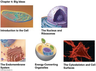
Introduction to the Cell
Historical Development and Importance of Microscopy
The development of microscopy revolutionized our understanding of cells, the fundamental units of life. Early scientists such as Robert Hooke and Anton van Leeuwenhoek made key discoveries using primitive microscopes, leading to the formulation of cell theory. Modern microscopy continues to expand our knowledge of cellular structure and function.
Robert Hooke (1665): First observed cells in cork using a compound microscope.
Anton van Leeuwenhoek (early 1700s): Used a simple microscope to observe living microorganisms in pond water and bacteria.
Matthias Schleiden (1838): Proposed that all plants are made of cells.
Theodor Schwann (1839): Proposed that all animals are made of cells.
Rudolf Virchow (1855): Stated that all cells arise from pre-existing cells.
Cell Theory:
All living things are composed of cells.
All cells come from other cells.
The cell is the basic unit of structure and function in organisms.
Microscopy and Cell Structure
Types of Microscopes
Microscopes are essential tools for studying cells. Different types of microscopes provide varying levels of magnification and resolution, allowing scientists to observe cellular structures in detail.
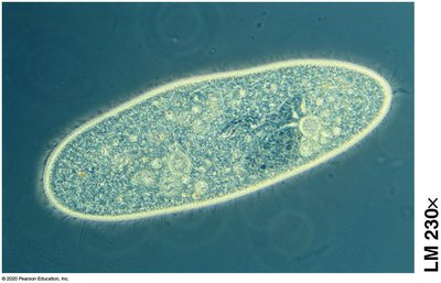
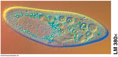
Light Microscope (LM): Uses visible light to illuminate specimens; can observe living cells and tissues.
Electron Microscopes (EM): Use beams of electrons for much higher resolution and magnification.
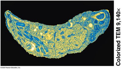
Transmission Electron Microscope (TEM): Reveals internal cell structures by passing electrons through thin sections of specimens.
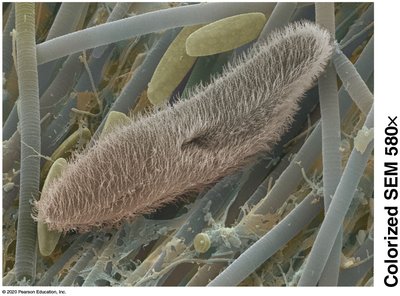
Scanning Electron Microscope (SEM): Provides detailed images of cell surfaces by scanning the specimen with electrons.
Key Terms:
Magnification: The increase in an object's image size compared to its actual size.
Resolution: The clarity of an image; the ability to distinguish two close objects as separate.
Microscope Type | Main Use | Example Image |
|---|---|---|
Light Microscope (LM) | Living cells, general structure |
|
Scanning Electron Microscope (SEM) | Surface details |
|
Transmission Electron Microscope (TEM) | Internal structures |
|
Light Microscope with Fluorescent Stain | Specific molecules/structures |
|
Cell Size and Surface Area
Surface-to-Volume Ratio
The small size of cells is crucial for efficient exchange of materials with their environment. A high surface-to-volume ratio allows for more effective transport of nutrients, gases, and waste products across the plasma membrane.
Plasma Membrane: A phospholipid bilayer with embedded proteins that regulates the movement of substances in and out of the cell.
Proteins in the membrane may act as channels or pumps for specific molecules.
Formula for Surface Area to Volume Ratio (for a cube):
As cell size increases, the surface area to volume ratio decreases, limiting the efficiency of exchange.
Prokaryotic vs. Eukaryotic Cells
Basic Features of All Cells
Plasma membrane
DNA (genetic material)
Ribosomes (protein synthesis)
Cytosol (fluid interior)
Prokaryotic Cells
Domains: Bacteria and Archaea
No membrane-bound nucleus or organelles
Generally smaller and simpler
Eukaryotic Cells
Domain: Eukarya (includes plants, animals, fungi, protists)
Membrane-bound nucleus
Numerous membrane-bound organelles
Compartmentalization in Eukaryotic Cells
Functional Groups of Organelles
Eukaryotic cells are organized into compartments, each with specialized functions:
Genetic Control: Nucleus and ribosomes
Manufacture, Distribution, Breakdown: Endoplasmic reticulum, Golgi apparatus, lysosomes, vacuoles, peroxisomes
Energy Processing: Mitochondria (all cells), chloroplasts (plants and algae)
Structural Support, Movement, Communication: Cytoskeleton, plasma membrane, cell wall (plants)
The Nucleus and Ribosomes
Nucleus
Contains the cell's DNA, which directs protein synthesis via messenger RNA (mRNA).
The nucleolus within the nucleus assembles ribosome subunits.
Ribosomes
Composed of ribosomal RNA and proteins.
Sites of protein synthesis, following instructions from DNA.
Can be free in the cytosol or bound to the endoplasmic reticulum.
The Endomembrane System
Components and Functions
Endoplasmic Reticulum (ER): Network of membranes; smooth ER synthesizes lipids and detoxifies, rough ER has ribosomes and produces proteins.
Golgi Apparatus: Modifies, sorts, and ships products from the ER to other destinations.
Lysosomes: Contain digestive enzymes to break down macromolecules and recycle cell components.
Vacuoles: Large vesicles for storage and maintenance; central vacuole in plants stores water and nutrients.
Peroxisomes: Metabolic compartments that break down fatty acids and detoxify harmful substances (not part of the endomembrane system).
Energy-Converting Organelles
Mitochondria
Site of cellular respiration, converting chemical energy from food into ATP.
Structure includes an outer membrane, inner membrane, intermembrane space, and matrix (contains DNA, ribosomes, enzymes).
Chloroplasts
Found in plants and algae; site of photosynthesis (conversion of solar energy to chemical energy in sugars).
Contain internal membranes (thylakoids) where light reactions occur.
Endosymbiont Theory
Mitochondria and chloroplasts likely evolved from free-living prokaryotes that were engulfed by ancestral eukaryotic cells.
The Cytoskeleton and Cell Surfaces
Cytoskeleton
Network of protein fibers: microfilaments, intermediate filaments, microtubules.
Functions: maintain cell shape, anchor/move organelles, cell movement, muscle contraction.
Cilia and Flagella
Locomotor appendages made of microtubules in a "9+2" arrangement.
Flagella: long, whip-like; cilia: short, coordinated movement.
Extracellular Matrix (ECM) and Cell Junctions
ECM: Network outside animal cells for support, adhesion, and communication.
Cell Junctions:
Tight junctions: Seal cells together.
Anchoring junctions: Fasten cells into strong sheets.
Gap junctions: Allow communication between cells.
Plant Cell Walls and Plasmodesmata
Rigid cell wall (mainly cellulose) provides support and protection.
Plasmodesmata: Channels that connect plant cells for transport and communication.
Summary Table: Eukaryotic Cell Structures and Functions
Organelle/Structure | Main Function |
|---|---|
Nucleus | Genetic control, houses DNA |
Ribosomes | Protein synthesis |
Endoplasmic Reticulum (ER) | Lipid and protein synthesis, detoxification |
Golgi Apparatus | Modification and sorting of macromolecules |
Lysosomes | Digestion and recycling |
Vacuoles | Storage, waste disposal, growth (plants) |
Mitochondria | ATP production (cellular respiration) |
Chloroplasts | Photosynthesis (plants, algae) |
Cytoskeleton | Structural support, movement |
Plasma Membrane | Selective barrier, communication |
Cell Wall (plants) | Protection, support |
Key Concepts for Review
Microscopes are essential for studying cell structure and function.
Cell theory is foundational to biology.
Prokaryotic and eukaryotic cells differ in complexity and compartmentalization.
Cell size is limited by the surface area to volume ratio.
Membrane structure is critical for cellular function.
Compartmentalization allows eukaryotic cells to carry out specialized functions efficiently.
Plant and animal cells have both shared and unique structures.
Mitochondria and chloroplasts have evolutionary origins explained by endosymbiosis.
The cytoskeleton, ECM, and cell junctions are vital for structure, movement, and communication.
