 Indietro
A Tour of the Cell: Structure, Types, and Microscopy
Guida di studio - Note intelligenti
 Appunti personalizzati basati sui tuoi materiali, ampliati con definizioni chiave, esempi e contesto.
The Tools for Viewing Cells
Microscopy in Cell Biology
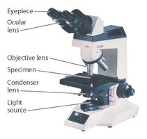
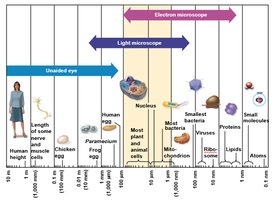
Microscopes are essential instruments in biology, allowing scientists to observe cells and their structures. There are two main types of microscopes used in cell biology: the light microscope (LM) and the electron microscope (EM).
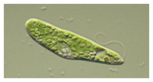
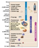
Light Microscope (LM): Uses visible light to magnify cells up to 1,000X, enabling observation of cell shape and structure.
Electron Microscope (EM): Uses electron beams for much higher magnification (up to 2 million X), revealing cellular details. However, EM cannot be used for living specimens.
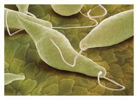
Scanning Electron Microscope (SEM): Provides detailed images of cell surface structures.
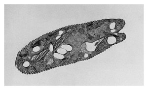
Transmission Electron Microscope (TEM): Reveals internal cell details.
The Cell Theory
Fundamental Principles of Cellular Life
The cell theory is a cornerstone of biology, stating that all living things are composed of cells, which are the smallest units of life. New cells arise from the division of pre-existing cells.
All living organisms are made of cells.
Cells are the basic unit of structure and function in living things.
Cells arise only from pre-existing cells.
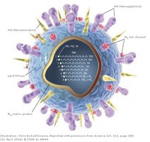
Viruses are not considered living: They lack cellular structure and cannot reproduce independently.
Relative Sizes of Cells
Why Cells Must Be Small
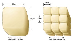
Cell size is limited by the surface area-to-volume ratio. Small cells have a higher ratio, allowing efficient exchange of nutrients and wastes with their environment. As cells grow larger, their volume increases faster than their surface area, reducing efficiency.
Surface Area: The area available for exchange of materials.
Volume: The space inside the cell where metabolic processes occur.
Surface Area-to-Volume Ratio: Determines how efficiently a cell can transport materials.
Characteristics of All Cells
Universal Features of Cells
Despite their diversity, all cells share certain fundamental characteristics:
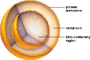
Plasma Membrane: A phospholipid bilayer that encloses the cell, controlling the movement of substances in and out.
Cytoplasm: The jelly-like substance inside the cell where organelles are suspended.
DNA: The genetic material that directs cell activities.
Ribosomes: Structures responsible for protein synthesis.
Types of Cells: Prokaryote vs. Eukaryote
Comparing Cell Types
Cells are classified into two main types: prokaryotic and eukaryotic. Prokaryotes are simpler and lack a nucleus, while eukaryotes have a nucleus and complex organelles.
Prokaryotic Cells: Found in bacteria and archaea. They lack a membrane-bound nucleus and organelles.
Eukaryotic Cells: Found in plants, animals, fungi, and protists. They have a nucleus and various membrane-bound organelles.
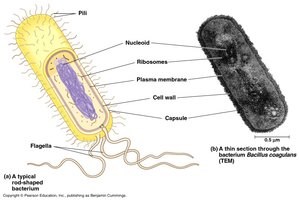
A Typical Bacterial (Prokaryotic) Cell
Bacterial cells have unique features such as a cell wall, capsule, pili, and flagella. The cell wall can be destroyed by antibiotics like penicillin. Capsules and pili aid in attachment to surfaces.
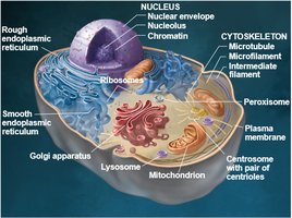
A Typical Animal (Eukaryotic) Cell
Animal cells contain a variety of organelles, each with specific functions. These include the nucleus, endoplasmic reticulum, Golgi apparatus, lysosomes, mitochondria, and cytoskeleton.
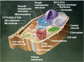
A Typical Plant Cell
Plant cells have additional organelles such as the cell wall, chloroplasts, and central vacuole, which are not found in animal cells.
Organelles of the Endomembrane System
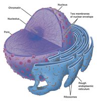
Nucleus
The nucleus is the largest organelle in eukaryotic cells, surrounded by a double-layered nuclear envelope with pores. It contains DNA and directs all cellular activities. The nucleolus within the nucleus is responsible for making ribosomes.
Nuclear Envelope: Double membrane with pores for material exchange.
Chromatin: DNA and associated proteins.
Nucleolus: Site of ribosome synthesis.
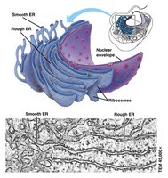
Smooth Endoplasmic Reticulum (ER)
The smooth ER synthesizes lipids, processes toxins and drugs, and stores/releases calcium ions in muscle cells.
Lipid Synthesis: Production of fats and steroids.
Detoxification: Breakdown of harmful substances.
Calcium Storage: Regulation of muscle contraction.
Summary Table: Prokaryotic vs. Eukaryotic Cells
Feature | Prokaryotic Cell | Eukaryotic Cell |
|---|---|---|
Nucleus | Absent | Present |
Membrane-bound Organelles | Absent | Present |
Cell Wall | Present (most) | Present (plants, fungi) |
Size | Small (1-10 µm) | Larger (10-100 µm) |
Examples | Bacteria, Archaea | Plants, Animals, Fungi, Protists |
Surface Area to Volume Ratio Formula
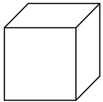
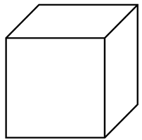
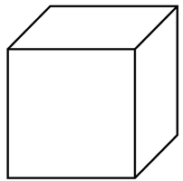
The surface area to volume ratio for a cube-shaped cell is calculated as:
Example: For a cube with side length 1 cm, the ratio is 6:1; for a cube with side length 3 cm, the ratio is 2:1.
Additional info:
These notes expand on the original content by providing definitions, examples, and formulas, ensuring a comprehensive and self-contained study guide for General Biology students.
