 Indietro
Bio 102 Week 7 Cancer A
Guida di studio - Note intelligenti
 Appunti personalizzati basati sui tuoi materiali, ampliati con definizioni chiave, esempi e contesto.
BIO 102 Week 7: Biology of Cancer
Overview of Cancer as a Biological Disease
Cancer is a complex disease characterized by uncontrolled cell division and the ability to adapt and evade normal regulatory mechanisms. It affects individuals and families worldwide, and remains a major challenge in medicine due to its diversity and adaptability. Understanding cancer requires integrating knowledge from cell biology, genetics, and physiology, as covered in foundational biology courses.
Hallmarks of Cancer Cells
Properties Contributing to Cancerous Growth
Cancer cells exhibit several distinct properties that differentiate them from normal cells. These hallmarks are essential for understanding cancer progression and diagnosis.
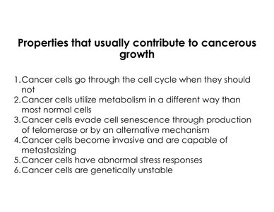
Uncontrolled Cell Cycle Progression: Cancer cells bypass normal cell cycle checkpoints and divide when they should not.
Altered Metabolism: Cancer cells utilize metabolic pathways differently, often favoring glycolysis even in the presence of oxygen.
Evasion of Cellular Senescence: Through production of telomerase or alternative mechanisms, cancer cells avoid the normal halt in division.
Invasiveness and Metastasis: Cancer cells can invade surrounding tissues and spread to distant sites.
Abnormal Stress Responses: Cancer cells respond differently to cellular stress, often avoiding programmed cell death.
Genetic Instability: Cancer cells accumulate mutations and chromosomal abnormalities at a higher rate.
Cell Cycle Regulation and Cancer
Cell Cycle Control Mechanisms
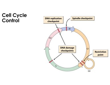
The cell cycle is tightly regulated by checkpoints that ensure DNA integrity and proper division. Cancer cells often ignore these controls, leading to abnormal proliferation and genetic instability.
Restriction Point (G1): Determines if a cell will proceed to division or enter a quiescent state (G0).
DNA Replication Checkpoint (S): Ensures DNA is properly replicated before division.
Spindle Checkpoint (M): Ensures chromosomes are correctly segregated during mitosis.
DNA Damage Checkpoints: Halt the cycle if DNA is damaged, allowing for repair or apoptosis.
Cyclins and Cyclin-Dependent Kinases (CDKs)
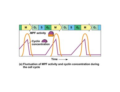
Cyclins are proteins that regulate the cell cycle by activating CDKs. The concentration of cyclins fluctuates throughout the cycle, controlling progression through different phases. Cancer cells may exploit these regulatory proteins to drive uncontrolled division.
MPF (Maturation Promoting Factor): A complex of cyclin and CDK that triggers mitosis.
Regulation: Peaks in cyclin concentration correspond to cell cycle transitions.
In Vitro Behavior of Cancer Cells
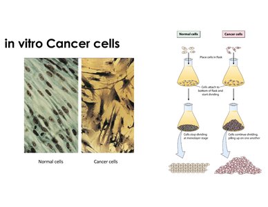
Normal cells exhibit anchorage dependence and density-dependent inhibition, stopping division when they reach confluence. Cancer cells lack these controls, continuing to divide and pile up even when crowded or detached from the substrate.
Anchorage Dependence: Normal cells require attachment to a substrate to survive and divide.
Density-Dependent Inhibition: Normal cells stop dividing when they form a monolayer.
Cancer Cells: Continue dividing beyond confluence, lack anchorage dependence, and display abnormal morphology.
Altered Metabolism in Cancer Cells
The Warburg Effect
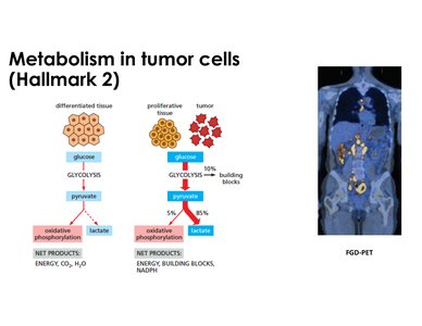
Cancer cells often rely on glycolysis for energy production, even in the presence of oxygen (aerobic glycolysis). This is less efficient than oxidative phosphorylation but supports rapid growth and provides precursors for biosynthesis.
Normal Cells: Use glycolysis followed by oxidative phosphorylation under aerobic conditions, producing more ATP per glucose.
Cancer Cells: Prefer glycolysis, producing lactate and requiring increased glucose uptake.
Warburg Effect: Named after Otto Warburg, describes this metabolic shift in cancer cells.
Clinical Application: Tumors can be imaged using PET scans with radio-labeled glucose due to their high uptake.
Telomere Maintenance and Cellular Immortality
Telomere Attrition and Telomerase
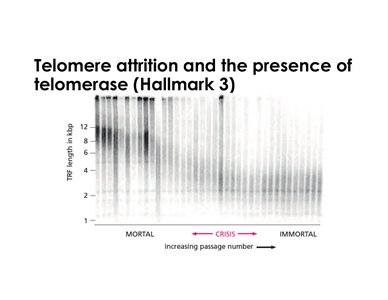
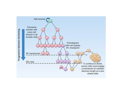
Telomeres are protective DNA sequences at chromosome ends that shorten with each division. When telomeres become critically short, cells enter senescence or crisis. Cancer cells evade this by expressing telomerase or using alternative mechanisms, allowing unlimited division.
Telomerase: An enzyme that extends telomeres, commonly reactivated in cancer cells.
Senescence: A state where cells stop dividing due to short telomeres.
Immortalization: Cancer cells maintain telomere length, avoiding senescence and crisis.
HeLa Cells: A famous example of immortalized cancer cells.
Alternative Lengthening of Telomeres (ALT)
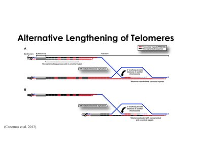
Some cancer cells use ALT, a telomerase-independent mechanism involving homologous recombination to maintain telomere length. This results in highly variable telomere lengths compared to telomerase-positive cells.
ALT Mechanism: Strand invasion and replication using telomeric repeats from other chromosomes.
Prevalence: About 15% of human tumors use ALT.
Comparison: Telomerase-positive cells have more consistent telomere lengths; ALT cells are heterogeneous.
Telomere Dynamics in Cell Populations
Stem cells and embryonic cells express telomerase to maintain telomere length. As cells differentiate, telomerase expression is lost, leading to telomere shortening and eventual senescence. Cancer cells bypass these checkpoints, maintaining telomere length through telomerase or ALT.
Self-Renewal: Stem cells maintain telomeres for continuous division.
Senescence and Crisis: Differentiated cells eventually stop dividing or die when telomeres are too short.
Cancer Cells: Bypass checkpoints and maintain telomere length, enabling unlimited proliferation.
Tumorigenesis: Benign vs. Malignant Tumors
Stages of Tumor Formation
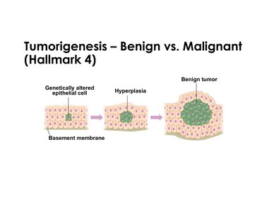
Tumorigenesis involves the transformation of normal cells into hyperplastic and then tumor cells. Benign tumors exhibit localized growth without invasion, while malignant tumors invade surrounding tissues and can metastasize.
Genetically Altered Cell: Initial mutation leads to abnormal proliferation.
Hyperplasia: Excessive but organized cell growth.
Benign Tumor: Localized, non-invasive growth.
Malignant Tumor: Invasive, poorly defined boundaries, capable of spreading.
Clinical Impact: Benign tumors are usually harmless; malignant tumors are responsible for most cancer-related deaths.
Comparison of Tumor Types
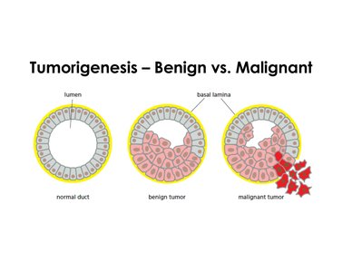
Benign and malignant tumors differ in morphology, invasiveness, and clinical outcomes. Malignant tumors break through the basal lamina and can migrate to other tissues, a process known as metastasis.
Normal Duct: Uniform cells, intact basal lamina.
Benign Tumor: Excess growth, regular nuclear shape, well-defined boundaries.
Malignant Tumor: Irregular nuclear shape, poorly defined boundaries, invasive behavior.
Summary Table: Hallmarks of Cancer Cells
Hallmark | Description | Example/Mechanism |
|---|---|---|
Uncontrolled Cell Cycle | Bypass of cell cycle checkpoints | Loss of restriction point control |
Altered Metabolism | Preference for glycolysis (Warburg effect) | Increased glucose uptake, lactate production |
Evasion of Senescence | Maintenance of telomere length | Telomerase expression or ALT |
Invasiveness/Metastasis | Ability to invade and migrate | Breakthrough basal lamina, metastasis |
Abnormal Stress Response | Failure to undergo apoptosis | Resistance to cell death signals |
Genetic Instability | Accumulation of mutations | Abnormal karyotypes, chromosomal changes |
