 Indietro
Biomolecules, Cell Components, and Membrane Structure: Study Notes for General Biology
Guida di studio - Note intelligenti
 Appunti personalizzati basati sui tuoi materiali, ampliati con definizioni chiave, esempi e contesto.
Biomolecules
Macromolecules and Polymers
Macromolecules are large biological molecules essential for life, composed of smaller building blocks. The four main types are proteins, carbohydrates, lipids, and nucleic acids. Polymers are macromolecules made of repeating units called monomers. Lipids are unique among macromolecules because they are not polymers; they are not made from repeating monomer units.
Macromolecules: Proteins, carbohydrates, lipids, nucleic acids
Polymers: Proteins, carbohydrates, nucleic acids
Lipids: Not polymers
Types of Carbohydrates
Carbohydrates are classified based on the number of sugar units:
Monosaccharides: Glucose, fructose, galactose, ribose
Disaccharides: Maltose (glucose + glucose), sucrose (glucose + fructose), lactose (glucose + galactose)
Polysaccharides: Starch, cellulose, glycogen, chitin
Formation and Function of Carbohydrates
Starch: Energy storage in plants, made of glucose
Cellulose: Structural support in plants, made of glucose (different linkage than starch)
Glycogen: Energy storage in animals, made of glucose
Chitin: Structural support in fungi and exoskeletons of insects, made of modified glucose with nitrogen
Comparison of Starch and Cellulose
Similarities: Both are made from glucose and found in plants
Differences: Different glucose linkages; starch is for energy storage, cellulose is for structure
Dehydration and Hydrolysis Reactions
Dehydration synthesis: Joins monomers by removing water
Hydrolysis: Breaks polymers by adding water
Covalent Bonds in Macromolecules
Proteins: Peptide bonds
Nucleic acids: Phosphodiester bonds
Carbohydrates: Glycosidic linkages
Lipids: Ester bonds
Lipids: Properties and Types
Properties: Hydrophobic, include fats and waxes
Phospholipids: Hydrophilic phosphate head, hydrophobic fatty acid tails
Triglycerides: Glycerol head, three fatty acid tails (saturated, unsaturated, trans fats)
Saturated vs. Unsaturated Fats
Saturated fats: Solid at room temperature, maximum hydrogen atoms, no double bonds
Unsaturated fats: Liquid at room temperature, fewer hydrogen atoms, one or more double bonds
Trans fats: Double bond in trans configuration, does not cause a kink
High unsaturated phospholipid concentration: Increases membrane fluidity
Structure of Polypeptides
Polypeptides: Chains of amino acids linked by peptide bonds
C-terminus: Carboxyl group end
N-terminus: Amino group end
Amino Acids: Structure and Properties
All amino acids have a carboxyl group, amino group, and hydrogen
Differences are in the R-group:
Polar: Contains oxygen or nitrogen
Nonpolar: Contains carbon or hydrogen
Acidic: Extra carboxyl group
Basic: Extra amino group
Protein Structure
Primary: Sequence of amino acids (peptide bonds)
Secondary: Hydrogen bonds form folds (alpha helix, beta sheet)
Tertiary: R-group interactions (hydrogen bonds, ionic bonds, hydrophobic interactions)
Quaternary: Multiple polypeptide chains assemble
Nucleic Acids: Purines and Pyrimidines
Purines: Adenine, guanine (double ring)
Pyrimidines: Thymine, cytosine, uracil (single ring)
Nucleotide Structure
Phosphate group
Nitrogenous base
Pentose sugar: Ribose or deoxyribose
DNA vs. RNA
DNA: Deoxyribose, thymine, double helix
RNA: Ribose, uracil, single helix
Base Pairing Rule in DNA
A-T
G-C
Cell Components
Animal vs. Plant Cells
Animal cells: Centrosomes, lysosomes
Plant cells: Large central vacuole, chloroplast, cell wall
Endomembrane System
Nuclear envelope
Endoplasmic reticulum (rough and smooth)
Golgi apparatus
Lysosomes
Vacuoles
Cell membrane
Protein Synthesis and Export
DNA transcribed to mRNA in nucleus
mRNA exits via nuclear pore
Ribosome on rough ER translates mRNA
Transport vesicle carries protein to Golgi
Golgi modifies and packages protein
Vesicle fuses with plasma membrane, releases protein
Prokaryotic vs. Eukaryotic Cells
Prokaryotes: No nucleus, no membrane-bound organelles, circular DNA, unicellular
Eukaryotes: Nucleus, membrane-bound organelles, linear DNA, uni/multicellular
Similarities Among All Cells
Ribosomes
Cytoplasm
Cell membrane
Genetic material
Free Ribosomes vs. Rough ER
Free ribosomes: Make proteins for use inside cell
Rough ER: Make proteins for secretion or membrane insertion
Functions of Eukaryotic Organelles
Nucleus: Stores DNA, control center
Mitochondria: ATP production
Rough ER: Protein synthesis
Smooth ER: Lipid synthesis, detoxification
Golgi apparatus: Modifies, sorts, packages molecules
Lysosomes: Digest waste, recycle organelles
Peroxisomes: Break down fatty acids, detoxify
Vacuoles: Storage, waste disposal, turgor pressure
Chloroplast: Photosynthesis
Ribosomes: Protein synthesis
Cytoskeleton: Shape, movement, anchor organelles
Centrosomes: Coordinate chromosome separation
Mitochondria and Chloroplasts
Contain their own DNA and ribosomes
Chromatin
DNA and histone proteins, involved in cell activities
Cell Junctions
Gap junctions: Communication in animal cells
Plasmodesmata: Communication in plant cells
Tight junctions: Leak-proof tissue in animals
Desmosomes: Hold tissues together in animals
Cytoskeletal Fibers
Microtubules: Tubulin, hollow tubes, cell shape, motility, chromosome movement
Microfilaments: Actin, intertwined strands, cell shape, motility, muscle contraction
Intermediate filaments: Keratin, coiled cables, cell shape, anchor organelles, nuclear lamina
Nuclear Lamina
Network of proteins supporting the nucleus
The Membrane
Plasma Membrane Structure and Selective Permeability
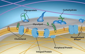
The plasma membrane is a dynamic structure composed of a phospholipid bilayer, proteins, carbohydrates, and cholesterol. It is selectively permeable, allowing only certain substances to pass through.
Phospholipids: Amphipathic molecules forming the bilayer
Proteins: Peripheral and integral, with various functions
Glycoproteins and glycolipids: Cell recognition
Cholesterol: Stabilizes membrane fluidity
Extracellular matrix (ECM): Collagen, fibronectin, cytoskeleton fibers
Lipids in the Plasma Membrane and Fluidity
Phospholipids: Amphipathic, form bilayer
Cholesterol: Maintains fluidity during temperature changes
Unsaturated fatty acids: Increase membrane fluidity
Extracellular Matrix and Plasma Membrane Proteins
ECM: Collagen fibers, fibronectin, cholesterol, cytoskeleton fibers
Plasma membrane proteins: Transport, enzymatic activity, signaling, cell recognition, intercellular joining, attachment
Transport Proteins
Channel proteins: Form tunnels, do not change shape (e.g., aquaporin)
Carrier proteins: Change shape to move molecules (e.g., sodium-potassium pump)
Integral vs. Peripheral Proteins
Integral proteins: Embedded in membrane, amphipathic
Peripheral proteins: Attached to membrane surface, hydrophilic
Glycoproteins and Glycolipids
Function in cell-to-cell recognition
Membrane Permeability
Small, hydrophobic molecules can easily cross
Diffusion and Transport
Simple diffusion: Movement from high to low concentration
Passive transport: No energy required
Facilitated diffusion: Uses transport proteins
Osmosis: Diffusion of water across a semipermeable membrane
Active Transport
Moves molecules from low to high concentration, uses ATP
Bulk transport uses vesicles and ATP
Endocytosis Types
Phagocytosis: Cell eating
Pinocytosis: Cell drinking
Receptor-mediated endocytosis: Specific uptake using membrane receptors
Tonicity and Effects on Cells
Hypotonic: Less solute outside
Hypertonic: More solute outside
Isotonic: Equal solute
Animal cells: Hypo - lysis, hyper - crenate, iso - normal
Plant cells: Hypo - turgid, hyper - plasmolyze, iso - flaccid
Turgor Pressure
Pressure of water inside plant cell pushing against cell wall
Normal Tonicity
Animal cells: Isotonic
Plant cells: Hypotonic
Membrane Potential
Difference in electrical charge across membrane
Maintained by ion channels and pumps (e.g., sodium-potassium pump, hydrogen pump)
Energy and Metabolism
Metabolic Pathways
Series of chemical reactions where product of one is substrate for next
Catabolic vs. Anabolic Pathways
Catabolic: Break down molecules, release energy
Anabolic: Build molecules, require energy
Spontaneous vs. Non-Spontaneous Processes
Spontaneous: No continuous energy input
Non-spontaneous: Requires energy input
Exergonic: Releases free energy
Endergonic: Requires free energy
Forms of Energy
Kinetic: Motion (e.g., moving car)
Potential: Stored (e.g., water behind dam)
Chemical: Bonds (e.g., glucose, ATP)
Thermal: Heat (e.g., hot water)
Light: Sunlight (e.g., photosynthesis)
Electrical: Charged particles (e.g., nerve signals)
Equilibrium and Free Energy
At equilibrium, ΔG = 0
Cells cannot do work at equilibrium
Exergonic and Endergonic Reactions
Exergonic: ΔG is negative, releases energy
Endergonic: ΔG is positive, requires energy
Laws of Thermodynamics
First law: Energy cannot be created or destroyed
Second law: Energy transfer increases entropy
Entropy
Measure of disorder; increases with energy transformations
Energy Coupling
Energy released by one reaction powers another
Example: ATP breakdown powers active transport
Enzymes and Activation Energy
Enzymes are proteins that speed up reactions by lowering activation energy
Do not change ΔG or get used up
Enzyme Structure and Specificity
Specific 3D shape
Active site binds substrate
Specificity due to shape and chemical properties
Optimal Temperature and pH
Enzymes work fastest at optimal temperature and pH
Enzyme Inhibitors
Competitive: Compete for active site
Non-competitive: Bind elsewhere, change enzyme shape
Cofactors and Coenzymes
Cofactors: Inorganic helpers (e.g., zinc, magnesium)
Coenzymes: Organic helpers (e.g., vitamins)
Key Equation: Free Energy Change
The change in free energy for a reaction is given by:
Where is the change in free energy, is the change in enthalpy, is the temperature in Kelvin, and is the change in entropy.
