Indietro
BIO 102 Week 8 Cancer Part 2 A
Guida di studio - Note intelligenti
 Appunti personalizzati basati sui tuoi materiali, ampliati con definizioni chiave, esempi e contesto.
BIO 102 Week 8 Cancer Part 2
Overview of Cancer Biology
Cancer is a complex disease involving multiple genetic and epigenetic changes that disrupt normal cellular processes. Understanding cancer requires integrating knowledge from cell cycle regulation, gene expression, signal transduction, and environmental influences. The progression from normal cells to malignant tumors is a multi-step process, often spanning decades, and involves both loss of function in tumor suppressor genes and gain of function in oncogenes.

Genetic and Epigenetic Changes in Cancer
Cancer Critical Genes: Tumor Suppressors and Oncogenes
Cancer progression is driven by mutations in two main classes of genes: tumor suppressor genes and proto-oncogenes. Tumor suppressors prevent inappropriate cell growth, while proto-oncogenes promote cell proliferation when activated. Mutations can lead to loss of function in tumor suppressors (e.g., APC, p53) or gain of function in oncogenes (e.g., Ras), resulting in uncontrolled cell division and tumor formation.
Tumor Suppressor Genes: Loss of function increases cancer risk. Example: APC mutation leads to colon polyps.
Oncogenes: Gain of function mutations convert proto-oncogenes to oncogenes. Example: Ras activation accelerates cell cycle progression.
Chromatin Accessibility: Changes in DNA methylation and histone modification alter gene expression, contributing to carcinogenesis.
Epigenetics and Cancer
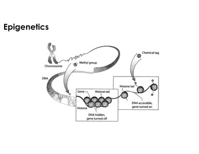
Epigenetics refers to heritable changes in gene expression that do not involve alterations in the DNA sequence. DNA methylation and histone modification are key epigenetic mechanisms. Hypermethylation typically silences genes, while hypomethylation can activate them. These changes affect chromatin structure, influencing accessibility for transcription and playing a significant role in cancer development.
DNA Methylation: Addition of methyl groups increases DNA compaction, reducing gene accessibility.
Histone Modification: Chemical tags on histone tails can relax chromatin, making genes accessible for transcription.
Gene Silencing: Hypermethylation of promoter regions can silence tumor suppressor genes.
Stem Cells and Cancer
Stem Cell Properties and Differentiation
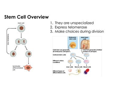
Stem cells are unspecialized cells with the ability to self-renew and differentiate into specialized cell types. They express telomerase, enabling extended replicative potential. During division, stem cells can either maintain their unspecialized state or commit to differentiation, leading to specialized cells.
Unspecialized: Stem cells lack a defined function until differentiation.
Telomerase Expression: Maintains chromosome ends, supporting longevity.
Potency: Embryonic stem cells are pluripotent (can form most cell types), while adult stem cells are multipotent (limited to tissue-specific cells).
Differentiation and Cancer Stem Cells
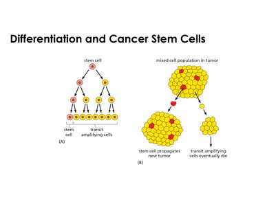
Cancer can arise from stem cells or through dedifferentiation of specialized cells. Two main theories explain cancer stem cell origin: dedifferentiation (reversion to a more plastic state) and the presence of cancer stem cells within tumors. Transit amplifying cells, derived from stem cells, proliferate rapidly and are often targeted by cancer treatments, but stem cells may evade therapy, leading to tumor recurrence.
Dedifferentiation: Terminally differentiated cells revert to a stem-like state, increasing plasticity and proliferation.
Cancer Stem Cells: Tumors may contain both stem cells and transit amplifying cells; stem cells are more resistant to treatment.
Therapeutic Implications: Targeting transit amplifying cells may reduce tumor size, but stem cells can regenerate the tumor.
Environmental and Chemical Influences on Cancer
Epidemiology and Cancer Risk
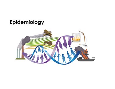
Epidemiology studies the distribution and determinants of diseases in populations. Environmental factors, such as pollution and lifestyle choices, significantly influence cancer incidence. Migration studies demonstrate that cancer rates can change with environmental exposure, highlighting the role of non-genetic factors in carcinogenesis.
Environmental Factors: Pollution, smoking, and diet can increase cancer risk.
Hereditary Factors: Genetic predisposition also contributes to cancer development.
Population Studies: Cancer rates vary geographically, reflecting environmental and lifestyle influences.
Sources of DNA Damage: Chemicals
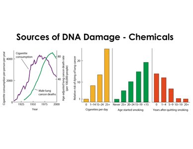
Chemicals such as those found in cigarette smoke are potent sources of DNA damage, leading to mutations and increased cancer risk. Epidemiological data show a strong correlation between cigarette consumption and lung cancer deaths, with risk increasing with the number of cigarettes smoked and the age at which smoking begins. Quitting smoking reduces cancer risk over time.
Cigarette Smoke: Contains mutagenic chemicals that damage DNA.
Risk Factors: Higher cigarette consumption and earlier age of initiation increase cancer risk.
Quitting Smoking: Reduces risk, demonstrating the impact of environmental modification.
Mutagenicity and Carcinogenicity
Ames Test: Assessing Mutagenicity
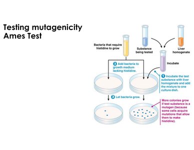
The Ames test is a laboratory assay used to determine whether a substance is a mutagen, which increases the likelihood of it being a carcinogen. The test uses bacteria that require histidine to grow; exposure to a mutagenic substance allows bacteria to acquire mutations enabling histidine synthesis, resulting in visible colonies on histidine-free media.
Mutagen: Substance that causes DNA mutations.
Carcinogen: Substance that causes cancer; most mutagens are carcinogens.
Experimental Design: Bacteria, test substance, and liver homogenate are incubated and plated; colony growth indicates mutagenicity.
Mutagenic and Carcinogenic Potency
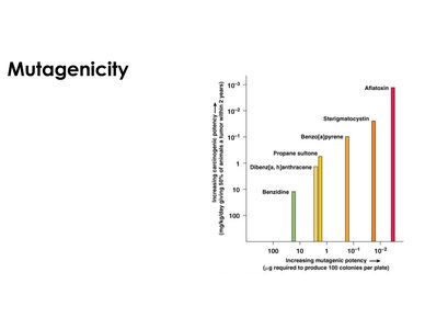
There is a direct relationship between mutagenic potency and carcinogenic potency. Substances requiring lower concentrations to produce mutations (as measured by the Ames test) are often more potent carcinogens. Potency is measured by the amount needed to induce mutations or tumors in experimental animals.
Mutagenic Potency: Measured by micrograms required to produce 100 colonies per plate.
Carcinogenic Potency: Measured by dose required to induce tumors in 50% of animals within two years.
Examples: Chemicals such as aflatoxin and benzidine are highly potent mutagens and carcinogens.
Cancer Critical Genes: Ras and p53
Ras: Proto-Oncogene and Oncogene
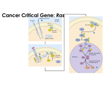
Ras is a GTPase involved in signal transduction pathways regulating cell proliferation. In its normal (proto-oncogene) state, Ras is activated by growth factor binding to enzyme-linked receptors, leading to downstream activation of cyclins and CDKs. A single point mutation can convert Ras into an oncogene, resulting in constitutive activation and uncontrolled cell division.
Normal Ras: Activated by GTP binding; promotes cell cycle progression.
Mutant Ras: Unable to hydrolyze GTP; remains active, driving excessive proliferation.
Oncogenic Impact: Dominant effect; only one mutated copy needed for cancer progression.
Receptor Abnormalities in Cancer
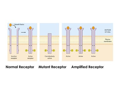
Cell surface receptors, especially those involved in growth factor signaling, can contribute to cancer when mutated or overexpressed. Mutant receptors may be constitutively active without ligand binding, while amplified receptors increase signaling in response to growth factors, both leading to excessive cell proliferation.
Normal Receptor: Activated by growth factor binding.
Mutant Receptor: Constitutively active, independent of growth factor.
Amplified Receptor: Overexpressed, increasing signal transduction.
p53: The Guardian of the Genome
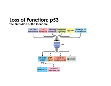
p53 is a tumor suppressor gene that regulates cell cycle arrest, DNA repair, apoptosis, and inhibition of angiogenesis in response to cellular stress. Loss of p53 function is common in many cancers, resulting in failure to control cell proliferation and repair DNA damage.
Activation: Triggered by stressors such as DNA damage, hypoxia, and radiation.
Function: Induces transcription of genes like p21 to inhibit CDKs and halt cell cycle progression.
Loss of Function: Leads to unchecked cell division and tumorigenesis.
Regulation of p53 Under Stress
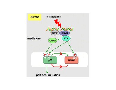
Under normal conditions, p53 is kept at low levels by MDM2-mediated degradation. Upon cellular stress (e.g., irradiation), p53 is phosphorylated, preventing its interaction with MDM2 and allowing p53 accumulation. This enables p53 to activate downstream mediators for cell cycle arrest, DNA repair, or apoptosis.
Phosphorylation: Prevents MDM2 inhibition, allowing p53 buildup.
Stress Response: Activation of p53 leads to protective cellular outcomes.
Mutations: Loss of both p53 copies impairs stress response and increases cancer risk.