Indietro
Bio 102 Week 7 Cancer B
Guida di studio - Note intelligenti
 Appunti personalizzati basati sui tuoi materiali, ampliati con definizioni chiave, esempi e contesto.
Metastasis: The Spread of Cancer Cells
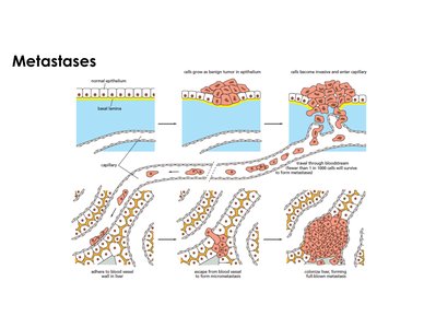
Metastasis is the process by which cancer cells spread from the primary tumor site to distant locations in the body, often leading to the formation of secondary tumors. This process is a hallmark of malignant cancers and is responsible for most cancer-related deaths.
Primary Tumor Formation: Cancer begins with abnormal cell proliferation, forming a benign tumor that does not invade surrounding tissues.
Transition to Malignancy: Malignant cells acquire the ability to invade through the basal lamina, entering blood or lymphatic vessels.
Circulatory Survival: Few cancer cells survive the journey through the bloodstream; fewer than 1 in 1000 cells successfully form metastases.
Extravasation and Colonization: Surviving cells exit microvessels in distant organs, forming micro-metastases, which may grow into macro-metastases.
Clinical Impact: Secondary tumors in vital organs (e.g., bone, lung, liver, brain) disrupt essential functions and are the primary cause of cancer mortality.
Metastatic Breast Cancer: Example and Clinical Relevance
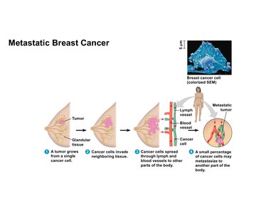
Breast cancer serves as a model for understanding metastasis. Early detection and localized treatment yield high survival rates, but metastatic spread significantly worsens prognosis.
Primary Tumor Location: Tumor originates in the mammary gland; removal of the gland can be curative if cancer is localized.
Invasion and Spread: Cancer cells invade neighboring tissue and enter blood or lymph vessels, enabling dissemination.
Secondary Sites: Metastases may occur in bone, lung, liver, or brain, causing severe physiological disruption.
Genetic Factors: Genes such as BRCA1 and BRCA2 act as tumor suppressors; mutations increase risk for breast and ovarian cancer.
Angiogenesis: Tumor-Induced Blood Vessel Formation
Mechanism and Importance of Angiogenesis
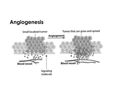
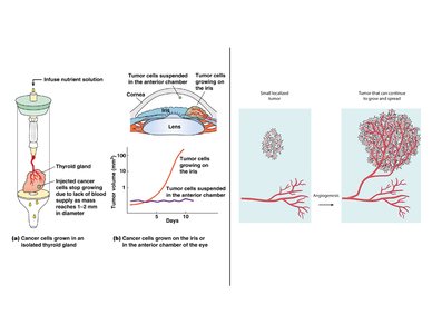
Angiogenesis is the formation of new blood vessels from pre-existing vasculature, a process exploited by tumors to ensure adequate oxygen and nutrient supply for continued growth and metastasis.
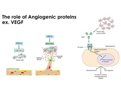
Hypoxic Environment: Rapidly proliferating tumor cells create regions of low oxygen (hypoxia), triggering angiogenesis.
Growth Factors: Tumor cells secrete angiogenic factors, notably VEGF (Vascular Endothelial Growth Factor), which stimulate endothelial cells to form new vessels.
Basal Lamina Degradation: Matrix metalloproteinases (MMPs) degrade the basal lamina, allowing vessel sprouting.
Fragile Vasculature: Newly formed vessels are permeable and fragile, facilitating tumor cell entry into circulation.
Clinical Note: Not all invasive tumors require angiogenesis; proximity to existing vessels may suffice.
Experimental Evidence for Angiogenesis
Experiments demonstrate the dependence of tumor growth on blood supply. Tumors lacking proximity to blood vessels remain small, while those near vessels grow rapidly.
In Vitro Assay: Cancer cells in isolated thyroid gland grow to 1-2 mm, then stop due to lack of blood supply.
In Vivo Assay: Tumor cells injected near the iris (highly vascular) expand significantly, while those in the anterior chamber (avascular) do not.
Necrosis vs. Apoptosis: Hypoxia leads to necrosis (cell death from lack of oxygen), distinct from apoptosis (programmed cell death).
Role of VEGF and Signal Transduction in Angiogenesis
VEGF is a key protein in angiogenesis, acting through signal transduction pathways to promote endothelial cell proliferation and migration.
Hypoxia-Inducible Factor (HIF-1α): Under low oxygen, HIF-1α increases, stimulating VEGF gene expression.
VEGF Gradient: Endothelial cells migrate toward areas of high VEGF concentration, forming capillary sprouts.
Balance of Factors: Cancer cells increase pro-angiogenic factors and decrease inhibitors, manipulating cell communication for their benefit.
Genetic Instability in Tumor Cells
Aneuploidy and Chromosomal Changes
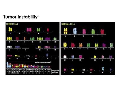
Tumor cells exhibit genetic instability, including aneuploidy (abnormal chromosome number), translocations, and increased heterochromatin, contributing to cancer progression and variability among patients.
Euploid vs. Aneuploid: Normal cells are euploid; tumor cells are often aneuploid.
Chromosomal Aberrations: Tumor cells may have extra chromosomes, losses, or translocations.
Heterochromatin: Increased compact chromatin is observed in cancer cells.
DNA Repair Deficiency: Ineffective repair pathways contribute to instability.
Cancer Critical Genes: Molecular Basis of Tumorigenesis
Definition and Categories
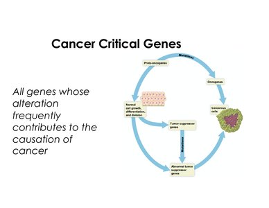
Cancer critical genes are those whose alteration frequently contributes to cancer. They fall into two main categories: proto-oncogenes (gain of function) and tumor suppressor genes (loss of function).
Proto-oncogenes: Normally promote cell growth and division; mutations convert them to oncogenes, driving uncontrolled proliferation.
Tumor Suppressor Genes: Normally inhibit inappropriate cell growth; mutations inactivate them, removing regulatory brakes.
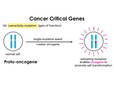
Gain of Function: Proto-oncogene to Oncogene
Gain of function mutations (overactivity) transform proto-oncogenes into oncogenes, promoting cell transformation and cancer. A single mutated copy is sufficient, indicating a dominant effect.
Mutation Types: Point mutations, gene amplification, chromosomal translocation, local DNA rearrangements, insertional mutagenesis.
Result: Hyperactive or overproduced proteins drive cell proliferation.
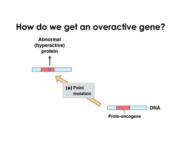
Point Mutation
Impact: Mutation in coding or regulatory region leads to hyperactive protein or overproduction of normal protein.
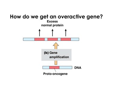
Gene Amplification
Impact: Multiple gene copies result in excess normal protein production.
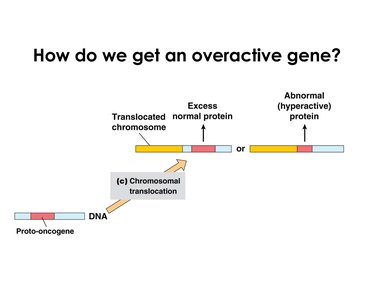
Chromosomal Translocation
Impact: Fusion proteins may be hyperactive; regulatory changes can cause overproduction of normal protein.
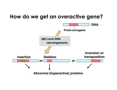
Local DNA Rearrangements
Impact: Insertion, deletion, inversion, or transposition can produce abnormal (hyperactive) proteins.
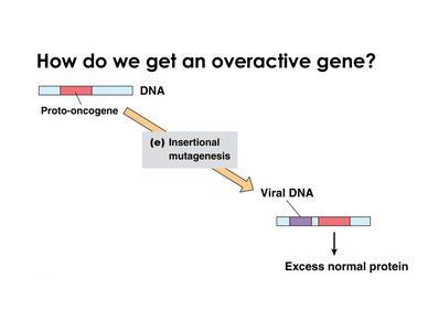
Insertional Mutagenesis
Impact: Viral DNA insertion near proto-oncogene deregulates expression, causing excess normal protein.
Loss of Function: Tumor Suppressor Genes
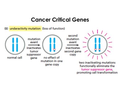
Loss of function mutations (underactivity) inactivate tumor suppressor genes. Both gene copies must be lost or mutated for cancer to develop, indicating a recessive effect.
Mutation Events: First mutation inactivates one copy; second mutation eliminates function, promoting cell transformation.
Mechanisms of Tumor Suppressor Loss
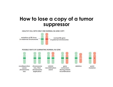
Non-disjunction: Chromosome loss during cell division.
Chromosome Duplication: After loss, duplication of mutated chromosome.
Mitotic Recombination: Exchange of genetic material during mitosis.
Gene Conversion: Conversion during recombination.
Deletion: Removal of normal gene copy.
Point Mutation: Inactivation of gene function.
Oncogenes vs. Tumor Suppressors: Functional Analogy
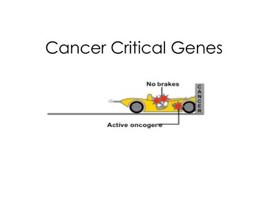
Oncogenes act as a gas pedal, accelerating cell proliferation, while loss of tumor suppressors removes the brakes, allowing uncontrolled growth. Both contribute to cancer progression.
Cancer as a Multistep Process
Stages of Cancer Development
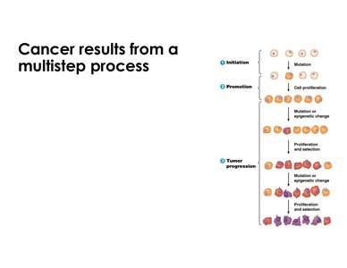
Cancer arises through a multistep process involving initiation, promotion, and progression. Multiple mutations and selective pressures are required for full transformation.
Initiation: Mutation converts cell to a precancerous state, increasing sensitivity to further changes.
Promotion: Repeated exposure to cancer-promoting agents leads to expansion of mutated cells.
Tumor Progression: Additional mutations and selection drive growth and differentiation of malignant cells.
Time Scale: The process can take years or decades, requiring multiple genetic and epigenetic changes.