 Indietro
Cell-Cell Interactions and Signaling in Plants and Animals
Guida di studio - Note intelligenti
 Appunti personalizzati basati sui tuoi materiali, ampliati con definizioni chiave, esempi e contesto.
Cell-Cell Interaction
Introduction to Cell-Cell Interaction
Cell-cell interaction is fundamental to multicellular life, enabling cells to communicate, coordinate responses, and maintain structural integrity. Cells are surrounded by extracellular materials that define their shape, attach them to other cells, and provide defense against external threats.
Extracellular structures are essential for cell shape and protection.
Cell communication is necessary for responding to environmental signals and molecule concentrations.
Cells secrete products that assemble into layers beyond the plasma membrane.
Extracellular Structures in Plant Cells
Organization of the Plant Cell Wall
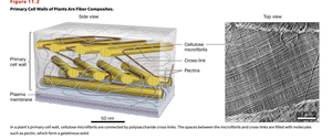
The plant cell wall is a fiber composite structure, crucial for plant cell shape and protection. It is composed mainly of cellulose microfibrils, cross-linked by polysaccharides, and filled with pectins.
Cellulose microfibrils provide structural support.
Pectins are hydrophilic polysaccharides that fill spaces between microfibrils, maintaining cell wall flexibility and hydration.
Microfibrils are synthesized directly into the extracellular space.
Pectins are synthesized in the rough ER and Golgi apparatus.
The primary cell wall maintains cell shape.
Turgor Pressure in Plant Cells
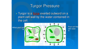
Turgor pressure is the force exerted outward on a plant cell wall by water contained in the cell. It is essential for maintaining cell rigidity and overall plant structure.
High solute concentration inside the cell causes water to enter via osmosis.
Incoming water increases cell volume, pushing the plasma membrane against the cell wall.
Turgor pressure is critical for plant cell function and growth.
Secondary Cell Walls in Plants
As plant cells mature, they may secrete a secondary cell wall, which varies in chemical composition depending on cell type and function.
Secondary cell walls may contain waxy coatings for waterproofing or lignin for rigidity.
Lignin is a polymer that makes cells sturdy and woody.
Secondary cell walls contain a high amount of cellulose.
Extracellular Matrix (ECM) in Animal Cells
Structure and Function of the ECM
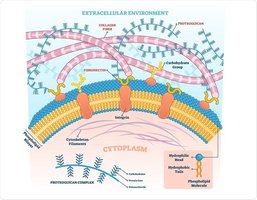
The ECM provides structural support to animal cells and contains more protein than carbohydrate. The main glycoprotein in the ECM is collagen, which gives tissues strength and elasticity.
Collagen is the primary structural protein in the ECM.
Proteoglycans are heavily glycosylated, gel-forming proteins responsible for the rubber-like consistency of cartilage.
ECM composition varies by tissue type.
Integrins and Laminins
Integrins are membrane proteins that bind to extracellular linking proteins called laminins, connecting the cytoskeleton to the ECM and facilitating cell adhesion and signaling.
Integrins and laminins are crucial for anchoring cells to the ECM.
They play a role in cell movement and signal transduction.
Cell-Cell Attachments
Indirect and Direct Cell-Cell Attachments
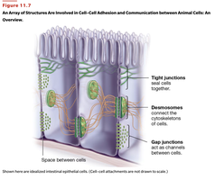
Cells connect to each other either indirectly via the ECM or directly through specialized junctions. These attachments are essential for tissue integrity and coordinated cellular functions.
Indirect attachments in animal cells occur through ECM components connected to membrane proteins like integrins.
Direct attachments include tight junctions, desmosomes, and gap junctions.
Tight Junctions
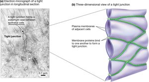
Tight junctions are found between cells forming barriers, such as epithelial cells lining the stomach and intestines. They create a watertight seal, preventing solutions from flowing between cells.
Tight junctions are dynamic and can loosen to permit certain ions.
They are weak adhesions and can be broken during cell wear and tear.
Desmosomes
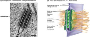
Desmosomes bind the cytoskeletons of cells together, providing strong adhesion in tissues such as muscle and epithelial cells. They connect to intracellular anchoring proteins and intermediate filaments.
Desmosomes act like rivets, holding cells together.
They are essential for tissue strength and integrity.
Cell-Cell Communication
Communication Between Adjacent Cells
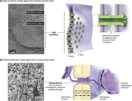
Cells in a tissue communicate through channels in their membranes, allowing diffusion of ions and small molecules. This exchange regulates gene expression and protein activity.
Signals can activate or deactivate proteins involved in metabolism, transport, secretion, and cytoskeleton.
Channels include gap junctions in animal cells and plasmodesmata in plant cells.
Plasmodesmata in Plant Cells
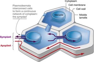
Plasmodesmata are channels that connect the cytoplasm of adjacent plant cells, forming a continuous network for communication and transport.
Plasmodesmata allow the flow of water, ions, and small molecules between cells.
They are essential for coordinated plant cell function.
Communication Between Distant Cells
Neurotransmitters and Hormones
Distant cell communication occurs via neurotransmitters and hormones. Neurotransmitters open and close ion channels in target cells, while hormones are information-carrying molecules that act on distant cells.
Hormones can be peptides, steroids, or gases.
They are secreted into bodily fluids and act on specific target cells.
Cell Signaling Pathways
General Steps in Cell Signaling
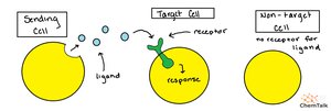
Cell signaling involves the release of a signal, binding to a receptor, transduction of the signal, and a cellular response. The process is dynamic and can be regulated by blocking receptors or altering signal transduction.
Signal molecules are produced or stored and released in response to conditions.
Receptors bind to signaling molecules and transduce the signal, often involving ATP.
The signaling molecule eventually unbinds, stopping the pathway.
Signal Reception and Processing
Receptors are dynamic and can be blocked by molecules such as beta blockers, which prevent adrenaline from binding to heart cell receptors.
Signal processing depends on the solubility of the signaling molecule.
Lipid-soluble signals are processed directly, while lipid-insoluble signals require transduction.
Lipid-Soluble Signal Processing
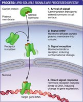
Lipid-soluble signals, such as steroid hormones, diffuse across the plasma membrane and bind to intracellular receptors, directly affecting gene expression.
Carrier proteins transport steroid hormones in the blood.
Hormones bind to receptors in the cytosol, inducing conformational changes.
The hormone-receptor complex binds to DNA, altering gene expression.
Lipid-Insoluble Signal Processing
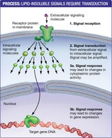
Lipid-insoluble signals bind to surface receptors, initiating a signal transduction cascade that generates intracellular signals and alters gene activity.
Signal transduction amplifies the signal and can lead to changes in cytoplasmic protein activity or gene expression.

G-Protein Coupled Receptors (GPCRs)
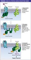
GPCRs are a major class of membrane receptors that activate intracellular signaling pathways via G-proteins. The process involves exchange of GDP for GTP, activation of enzymes, and production of second messengers.
Signal reception causes G-protein activation.
Activated G-protein triggers enzyme activity, producing second messengers.
Second Messengers
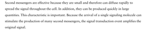
Second messengers are small molecules that rapidly diffuse and amplify the signal within the cell. They can be produced in large quantities and initiate diverse cellular responses.
Second messengers include calcium ions, cyclic AMP, cyclic GMP, diacylglycerol, and inositol triphosphate.
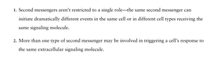
They are not restricted to a single role and can trigger different responses in different cell types.
Multiple second messengers may be involved in a single cellular response.

Name | Type of Response |
|---|---|
Calcium (Ca2+) | Binds to a protein called calmodulin; Ca2+-calmodulin complex then activates proteins |
Cyclic adenosine monophosphate (cAMP) | Activates certain protein kinases |
Cyclic guanosine monophosphate (cGMP) | Opens ion channels, activates certain protein kinases |
Diacylglycerol (DAG) | Activates certain protein kinases |
Inositol triphosphate (IP3) | Opens calcium channels, allowing stored calcium ions to enter cytosol |

Enzyme-Linked Receptors
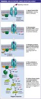
Enzyme-linked receptors, such as receptor tyrosine kinases, initiate phosphorylation cascades that amplify and regulate cellular responses. Phosphatases can terminate the signal by removing phosphate groups.
Signal reception activates the receptor's kinase activity.
Phosphorylation cascades amplify the signal.
Phosphatases remove phosphate groups, ending the signal transduction.
Summary Table: Types of Cell-Cell Attachments
Attachment Type | Location | Function |
|---|---|---|
Tight Junctions | Animal cells (epithelial tissues) | Seal cells together, prevent passage of molecules |
Desmosomes | Animal cells (muscle, epithelial tissues) | Bind cytoskeletons, provide mechanical strength |
Gap Junctions | Animal cells | Allow passage of ions and small molecules |
Plasmodesmata | Plant cells | Connect cytoplasm, allow communication and transport |
Conclusion
Cell-cell interactions and signaling are essential for multicellular life, enabling cells to communicate, coordinate activities, and maintain structural integrity. Understanding these processes is fundamental to biology and provides insight into how tissues and organisms function.
