 Indietro
Cell Communication and Cell-Cell Connections in Multicellular Organisms
Guida di studio - Note intelligenti
 Appunti personalizzati basati sui tuoi materiali, ampliati con definizioni chiave, esempi e contesto.
Cell Communication and Connections
Introduction to Cell Communication
Multicellular organisms rely on direct and indirect connections between cells to maintain structural integrity and coordinate cellular activities. These connections are essential for cells to determine their location, identity, and the behavior of neighboring cells.
Direct physical connections and extracellular matrix (ECM) interactions are fundamental to multicellularity.
Cell communication is necessary for development, tissue maintenance, and response to environmental signals.
Cell-Cell Connections in Plants
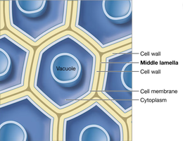
Plant cells are glued together by the middle lamella, a pectin-rich layer that provides structural cohesion.
The middle lamella is located between the cell walls of adjacent plant cells.
It enables plant tissues to withstand mechanical stress and maintain integrity.
Cell-Cell Connections in Animals
Animal cells lack cell walls but are anchored through the ECM and specialized membrane proteins.
Membrane proteins allow direct connections between adjacent cells.
ECM provides indirect anchoring and structural support.
Types of Direct Cell-Cell Connections in Animals
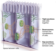
Animal cells use several types of junctions to connect and communicate:
Tight junctions: Form a waterproof seal, often found in epithelial cells. They regulate the passage of substances between cells.
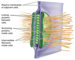
Desmosomes: Provide strong adhesion by connecting cytoskeletons of adjacent cells. They consist of linking proteins (cadherins) and anchoring proteins attached to intermediate filaments.
Gap junctions: Create channels for direct communication, allowing molecules and ions to pass between cells.
Tight Junctions
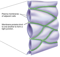
Tight junctions are formed by membrane proteins binding together, sealing cells and preventing leakage of extracellular fluid.
They are dynamic and can loosen or tighten depending on physiological needs.
Desmosomes
Desmosomes are composed of cadherins (linking proteins) and anchoring proteins, which connect to intermediate filaments inside cells.
They provide continuous structural support across tissues.
Cadherins are responsible for selective adhesion, ensuring cells of the same tissue type adhere together.
Selective Cell Adhesion
Selective adhesion is mediated by specific membrane proteins, such as cadherins.
Cadherins ensure that cells destined for the same tissue aggregate together during development.
Disruption of cadherin expression can lead to abnormal tissue formation.
Cell-Cell Communication via Gaps
Gap junctions (animals) and plasmodesmata (plants) allow adjacent cells to communicate by creating shared channels.
Signals can move through these channels to regulate gene expression or activate/inactivate proteins in neighboring cells.
Regulation of gene expression is typically slower, while direct protein activation is faster.
Signal Transduction and Cell Fate
Signal transduction is the process by which cells convert extracellular signals into intracellular responses.
Signals can travel between distant cells or require direct contact (e.g., Notch signaling).
Notch signaling is crucial for determining cell fate during development and relies on physical contact between adjacent cells.
Experimental Evidence for Cell Adhesion and Communication
Adhesion Proteins in Animal Cells
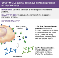
Experiments demonstrate that selective adhesion is due to specific membrane proteins.
Membrane proteins are isolated from cells that adhere to others of the same type.
Antibodies are produced to target these proteins, confirming their role in cell adhesion.
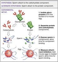
Role of Glycoproteins in Sperm-Egg Attachment
Glycoproteins on egg surfaces are composed of carbohydrate and protein components. Experiments test whether sperm attach to the carbohydrate or protein part.
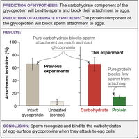
Carbohydrate component blocks sperm attachment as effectively as intact glycoprotein.
Protein component blocks few sperm from attaching.
Conclusion: Sperm recognize and bind to carbohydrates of egg-surface glycoproteins.
Summary Table: Types of Cell-Cell Connections
Type | Structure | Function | Location |
|---|---|---|---|
Tight Junction | Membrane proteins binding adjacent cells | Seals cells, prevents leakage | Epithelial tissues |
Desmosome | Cadherins, anchoring proteins, intermediate filaments | Strong adhesion, structural support | Animal tissues |
Gap Junction | Channel proteins | Direct communication, molecule transfer | Animal tissues |
Plasmodesmata | Channels through cell walls | Direct communication, molecule transfer | Plant tissues |
Middle Lamella | Pectin-rich layer | Glues plant cells together | Plant tissues |
Key Terms and Concepts
Cadherins: Membrane proteins responsible for selective cell adhesion.
Desmosomes: Junctions providing strong adhesion between animal cells.
Tight Junctions: Junctions sealing cells to prevent passage of substances.
Gap Junctions: Channels allowing communication between animal cells.
Plasmodesmata: Channels allowing communication between plant cells.
Middle Lamella: Layer gluing plant cells together.
Signal Transduction: Conversion of extracellular signals to intracellular responses.
Notch Signaling: Pathway determining cell fate via direct cell contact.
Example Application
During embryonic development, selective expression of cadherins ensures proper tissue formation.
Disruption of cell-cell junctions can lead to diseases such as cancer or tissue degeneration.
Equations and Academic Context
Cell adhesion strength can be quantified experimentally by measuring attachment inhibition percentage.
Signal transduction pathways often involve phosphorylation cascades, represented as:
Additional info: Academic context was added to clarify the role of cell junctions, signal transduction, and experimental evidence for cell adhesion.
