 Indietro
Cell Communication and Signal Transduction in Biology
Guida di studio - Note intelligenti
 Appunti personalizzati basati sui tuoi materiali, ampliati con definizioni chiave, esempi e contesto.
Cell Communication
Overview of Cellular Messaging
Cell-to-cell communication is fundamental for both multicellular and unicellular organisms, enabling regulation of cellular activities and responses to environmental cues. Cells primarily communicate via chemical signals, which can be transmitted over varying distances.
Internal and External Signals: Control cellular activity and coordinate responses.
Methods of Communication: Universal and unique mechanisms exist for cellular regulation.
Signaling Distances
Cell signaling can be classified based on the distance over which signals are transmitted:
Cell Contact: Direct communication via gap junctions (animal cells) or plasmodesmata (plant cells).
Local Signaling: Paracrine signaling and synaptic signaling involve local regulators and neurotransmitters.
Long Distance Signaling: Endocrine signaling uses hormones that travel through the bloodstream to distant target cells.
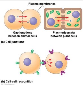
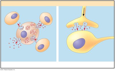
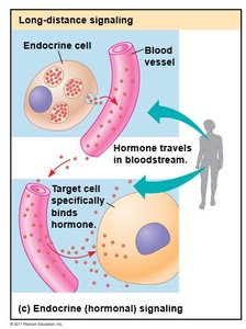
The Three Stages of Cell Signaling
Reception
Reception is the first step in cell signaling, where a cell detects a signaling molecule (ligand) from outside.
Ligand: The external signal molecule, binds specifically to a receptor (lock and key model).
Receptor: Usually located on the plasma membrane, but can also be intracellular.
Types of Receptors:
G protein-coupled receptors (GPCRs)
Receptor tyrosine kinases (RTKs)
Ion channel receptors
G Protein-Coupled Receptors (GPCRs)
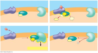
GPCRs are the largest family of cell-surface receptors, characterized by seven transmembrane helices. They interact with G proteins and GTP to transmit signals. 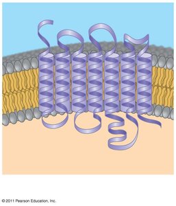
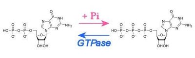
Activation: Ligand binding causes a conformational change, activating the G protein by exchanging GDP for GTP.
GTPase: Removes a phosphate group from GTP, turning the G protein off.
Receptor Tyrosine Kinases (RTKs)
RTKs are membrane receptors that catalyze the transfer of phosphate groups from ATP to tyrosines.
Dimerization: RTKs pair up upon ligand binding.
Phosphorylation: Tyrosines are phosphorylated, creating binding sites for relay proteins.
Multiple Pathways: RTKs can trigger several signal transduction pathways simultaneously.
Ligand-Gated Ion Channels
These receptors act as gates that open to allow ions (e.g., Na+, Ca2+) through when bound to a ligand.
Vital for Neurons: Essential for nerve impulse transmission and physiological processes.
Intracellular Receptors
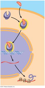
Found in the cytosol or nucleus, these receptors are activated by small or hydrophobic messengers (e.g., steroid hormones).
Example: Testosterone binds to the androgen receptor, which then regulates gene expression.
Transduction
Signal Transduction Pathways
Transduction involves cascades of molecular interactions that relay signals from receptors to target molecules.
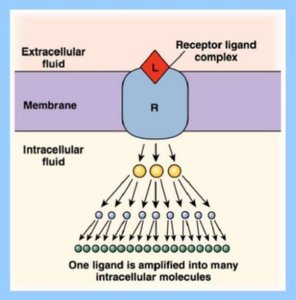
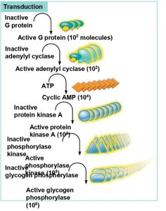
Amplification: Multistep pathways amplify the signal, allowing a few molecules to produce a large response.
Coordination: Multiple steps provide opportunities for regulation.
Phosphorylation Cascades
Protein kinases transfer phosphates from ATP to proteins (phosphorylation), while protein phosphatases remove them (dephosphorylation). This acts as a molecular switch. 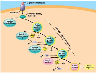
Second Messengers
Second messengers are small, non-protein, water-soluble molecules or ions that spread throughout the cell by diffusion.
Common Second Messengers: Cyclic AMP (cAMP) and calcium ions (Ca2+).
Cyclic AMP (cAMP)
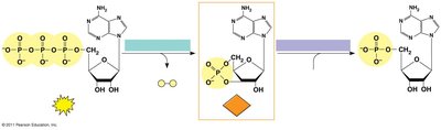
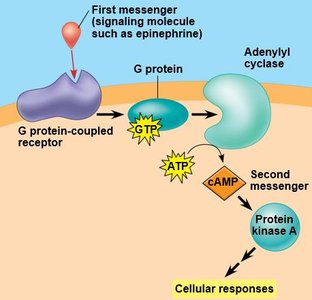
Adenylyl Cyclase: Converts ATP to cAMP in response to extracellular signals.
cAMP Pathway: Activates protein kinase A (PKA), which phosphorylates other proteins.
Calcium Ions (Ca2+)
Calcium ions act as second messengers in many pathways, with cells regulating their concentration internally. 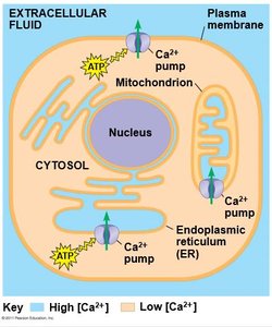
IP3 and Calcium Release
Inositol triphosphate (IP3) can activate ligand-gated calcium channels, triggering further transduction pathways. 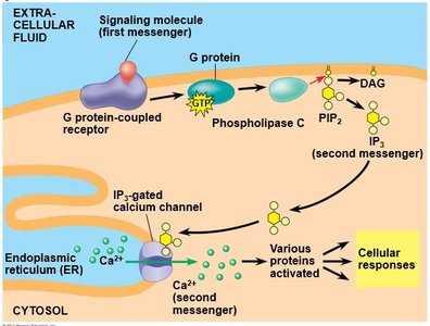
Response
Cellular Responses to Signal Transduction
The final stage of cell signaling leads to regulation of cellular activities, which may occur in the cytoplasm or nucleus.
Gene Expression: Many pathways regulate synthesis of enzymes or proteins by turning genes on or off.
Transcription Factors: The final activated molecule may act as a transcription factor.
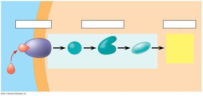
Fine-Tuning Cellular Responses
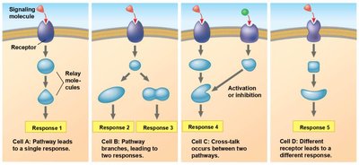
Four aspects of fine-tuning include:
Amplification: Enzyme cascades amplify the response.
Specificity: Different cells have unique protein collections, leading to specific responses.
Efficiency: Scaffolding proteins enhance signal transduction efficiency.
Termination: Inactivation mechanisms ensure signals are not perpetuated indefinitely.
Apoptosis: Programmed Cell Death
Triggering Apoptosis
Apoptosis is a rapid, irreversible process designed to remove damaged or unneeded cells.
Triggers: Irreversible DNA damage, severe signaling imbalances, or signals from other cells.
Developmental Role: Apoptosis sculpts features and maintains tissue health.
p53 Regulation of Cell Health
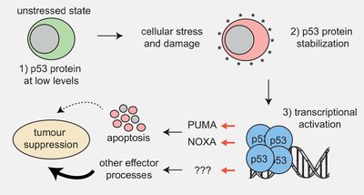
p53: Normally degraded, but rises in response to stress. Phosphorylated p53 acts as a transcription regulator.
The Apoptotic Program
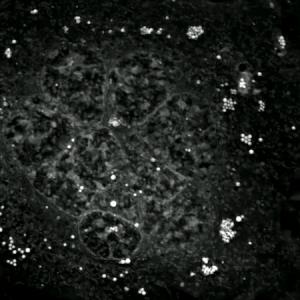
Apoptosis involves a series of organized steps:
Bleb formation
Nucleus collapse (pyknosis)
Chromosomal DNA cleavage
Organelle localization changes
Formation of apoptotic bodies
Mitochondria in Apoptosis
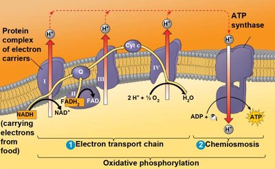
Mitochondria release cytochrome C during apoptosis, which triggers apoptotic pathways.
Case Study: Cancer Resistance in Elephants
Background
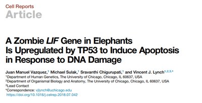
Larger animals, such as elephants, have more cells and should theoretically have higher cancer rates, but elephants have a much lower rate of cancer than humans.
LIF Gene: Leukemia inhibitory factor (LIF) is involved in preventing tumor formation by triggering apoptosis in response to DNA damage.
Elephants: Have multiple copies of the LIF gene, which may contribute to their cancer resistance.
Implications for Human Cancer Treatment
Increased LIF expression triggers apoptosis, suggesting potential strategies for cancer therapy.
Future Study: Investigating whether increasing LIF gene copies or expression in humans could reduce cancer rates.
Summary Table: Membrane Bound Receptors and Signal Transduction
Receptor | Notes | Signal Transduction | Notes |
|---|---|---|---|
GPCR | Uses G-protein + GTP/GDP | cAMP turns on PKA; cAMP made by adenylyl cyclase; IP3 opens calcium channels | Changes in Ca2+ concentration regulate multiple pathways |
RTK | Requires dimerization; works as a kinase | Phosphorylation of tyrosines | Triggers multiple pathways |
Ligand-gated ion channels | Opens ion channels | Ion flow regulates cell activity | Vital for neurons |
Key Terms and Concepts
Ligand: A molecule that binds to a receptor to initiate a signal.
Receptor: Protein that receives and transmits signals.
Kinase: Enzyme that transfers phosphate groups.
Phosphorylation: Addition of a phosphate group to a protein.
Second Messenger: Small molecule that amplifies and transmits signals inside the cell.
Apoptosis: Programmed cell death.
p53: Tumor suppressor protein regulating cell health and apoptosis.
Additional info: This guide expands on the original notes with academic context, definitions, and examples to ensure completeness and clarity for exam preparation.
