 Indietro
Cell Communication and Signal Transduction: Mechanisms and Pathways
Guida di studio - Note intelligenti
 Appunti personalizzati basati sui tuoi materiali, ampliati con definizioni chiave, esempi e contesto.
Cell Communication
Introduction to Cell Communication
Cell communication is essential for the coordination of cellular activities in both unicellular and multicellular organisms. Cells respond to external messengers through various mechanisms, enabling processes such as growth, development, immune responses, and homeostasis.
Cell signaling involves the transmission of information from one cell to another via signaling molecules.
Signaling mechanisms evolved in prokaryotes and single-celled eukaryotes and were adapted by multicellular organisms.
Evolution of Cell Signaling
The ability to communicate via signaling molecules is ancient and fundamental to life.
Prokaryotes and eukaryotes use ancestral signaling molecules for communication.
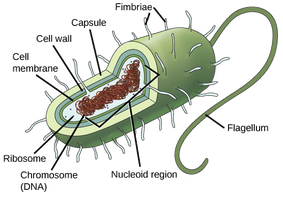
Quorum sensing in bacteria allows them to sense population density and coordinate behaviors such as biofilm formation and toxin secretion.
Interfering with quorum sensing is a potential strategy for combating bacterial infections.
Cell Signaling in Yeast
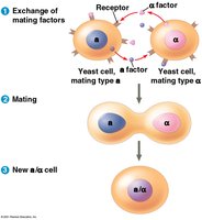
The yeast Saccharomyces cerevisiae has two mating types, a and α, which locate each other via secreted factors.
Binding of a mating factor initiates a signal transduction pathway.
Molecular details of signal transduction are conserved across yeasts and mammals.
Local and Long-Distance Signaling
Local Signaling
Cells in multicellular organisms communicate via signaling molecules that act locally or over long distances.
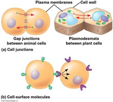
Direct contact: Animal and plant cells have cell junctions (gap junctions in animals, plasmodesmata in plants) that allow direct cytoplasmic exchange.
Cell-surface molecules: Cells may communicate by direct contact between cell-surface molecules, important in development and immune responses.
Paracrine and Synaptic Signaling
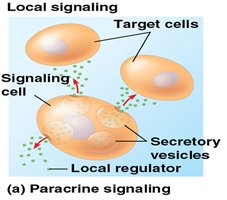
Paracrine signaling: Secreted messenger molecules travel short distances to stimulate nearby target cells (e.g., growth factors).
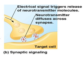
Synaptic signaling: Neurotransmitters are released in response to electrical signals and diffuse across synapses in the nervous system.
Long-Distance Signaling
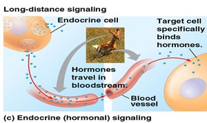
Hormonal (endocrine) signaling: Specialized cells release hormones that travel via the circulatory system to distant target cells.
The ability of a cell to respond depends on the presence of specific receptors for the signal.
The Three Stages of Cell Signaling
Overview of Cell Signaling
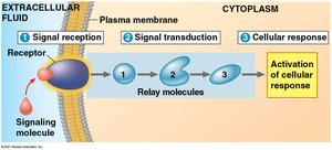
Earl W. Sutherland's research revealed that cells receiving signals undergo three main processes:
Signal Reception: Detection of a signaling molecule by a receptor protein on the cell surface.
Signal Transduction: The receptor initiates a signal transduction pathway, often involving a series of steps.
Cellular Response: The transduced signal triggers a specific response in the target cell.
Reception: Signal Molecule Binding
Mechanism of Reception
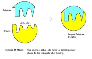
Signal molecules (ligands) bind to highly specific receptor proteins, causing a shape change that initiates signal transduction.
Most receptors are plasma membrane proteins, but some are located inside the cell.
Types of Membrane Receptors
There are three main types of membrane receptors:
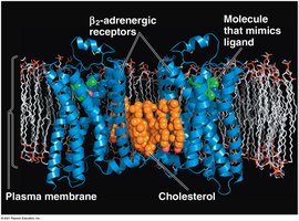
G protein-coupled receptors (GPCRs): Largest family of cell-surface receptors; work with G proteins that bind GTP.
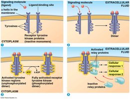
Receptor tyrosine kinases (RTKs): Catalyze transfer of phosphate groups from ATP to proteins; can trigger multiple pathways.
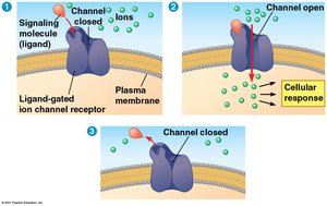
Ion channel receptors: Act as gates that open or close to allow specific ions through when the receptor changes shape.
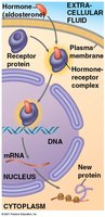
Intracellular Receptors
Located in the cytoplasm or nucleus of target cells.
Small or hydrophobic messengers (e.g., steroid and thyroid hormones) can cross the membrane and activate these receptors.
The hormone-receptor complex acts as a transcription factor, regulating gene expression.
Transduction: Signal Relay and Amplification
Signal Transduction Pathways
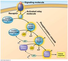
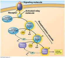
Cell signaling is usually a multistep process, with each step involving shape changes in proteins.
The signal is relayed and often amplified through a cascade of molecular interactions.
Protein Phosphorylation and Dephosphorylation
Protein kinases transfer phosphates from ATP to proteins (phosphorylation), creating a phosphorylation cascade.
Protein phosphatases remove phosphates (dephosphorylation), acting as molecular switches.
Second Messengers
Small, non-protein, water-soluble molecules or ions that spread by diffusion.
Common second messengers: cyclic AMP (cAMP) and calcium ions (Ca2+).
Cyclic AMP (cAMP) Pathway
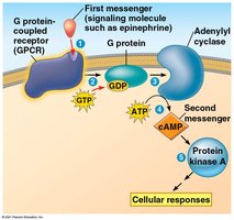
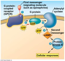
cAMP is produced from ATP by adenylyl cyclase in response to extracellular signals.
cAMP activates protein kinase A, which phosphorylates various proteins.
G protein systems can regulate adenylyl cyclase activity.

cAMP in Disease
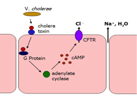
The cholera bacterium (Vibrio cholerae) produces a toxin that keeps G protein active, causing continuous cAMP production.
This leads to excessive salt and water secretion in the intestines, resulting in dehydration.
Calcium Ions and Inositol Triphosphate (IP3)
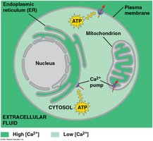
Ca2+ is a widely used second messenger; its cytosolic concentration is tightly regulated.
IP3 and diacylglycerol (DAG) are produced by cleavage of membrane phospholipids and help release Ca2+ from intracellular stores.
Cellular Responses
Nuclear and Cytoplasmic Responses
Signal transduction pathways regulate cellular activities in the nucleus or cytoplasm.
Many pathways regulate gene expression by activating transcription factors.
Other pathways regulate protein activity, such as opening ion channels or modifying metabolic enzymes.
Signal Amplification
Enzyme cascades amplify the cell’s response; each step increases the number of activated molecules.
Regulation of the Response
Aspects of Signal Regulation
Amplification: Signal and response are amplified through cascades.
Specificity: Different cells respond differently to the same signal due to unique protein collections.
Efficiency: Scaffolding proteins enhance signal transduction by grouping pathway components.
Termination: Inactivation mechanisms ensure signals are not perpetuated when external signals decrease.
Apoptosis: Programmed Cell Death
Mechanism and Importance of Apoptosis
Apoptosis is a controlled process where cells are dismantled and removed without harming neighboring cells.
Triggered by signals that activate a cascade of “suicide” proteins (caspases).
Essential for development, maintenance, and disease prevention in animals.
Apoptosis in Caenorhabditis elegans
Ced-9 protein acts as a brake; when inactivated, apoptosis proceeds via activation of proteases and nucleases (e.g., Ced-3).
Apoptosis in Mammals
Multiple pathways and caspases are involved; can be triggered by internal or external signals.
Normal in development (e.g., separation of fingers in embryos) and may be involved in diseases such as Parkinson’s and Alzheimer’s.
Additional info: Apoptosis is a highly regulated process, and its dysregulation can lead to cancer or degenerative diseases. ----------------------------------------
