Indietro
Cell Communication: Mechanisms and Examples
Guida di studio - Note intelligenti
 Appunti personalizzati basati sui tuoi materiali, ampliati con definizioni chiave, esempi e contesto.
Cell Communication
Overview of Cell Signaling
Cell communication is essential for multicellular life, enabling cells to coordinate activities and respond to environmental cues. Signals can be transmitted through direct contact, local signaling, or hormones. - Direct contact: Cells connect via gap junctions (animals) or plasmodesmata (plants) for molecule exchange. Tight junctions and desmosomes provide structural integrity. - Local signaling: Paracrine signaling involves nearby cells (e.g., growth factors), while synaptic signaling uses neurotransmitters across synapses. - Endocrine signaling: Hormones travel long distances via the bloodstream to target cells with specific receptors. 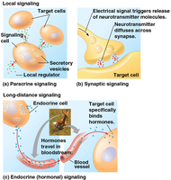
Hormones and Receptors
Hormones are signaling ligands that bind to specific receptors on target cells, triggering a cellular response. There are two major classes: - Lipid-soluble hormones: Cross the cell membrane and bind to intracellular receptors. - Lipid-insoluble hormones: Bind to cell-surface receptors, unable to cross the membrane.
Stages of Cell Signaling
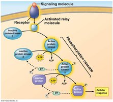
Cell signaling typically follows three stages: reception, transduction, and response. - Reception: The cell detects a signaling molecule from the outside. - Transduction: The signal is relayed and amplified inside the cell, often through a cascade of molecules. - Response: The cell reacts, such as by altering metabolism, gene expression, or cytoskeletal arrangement. 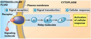
Signal Transduction Pathways
Example: Glycogen Breakdown by Epinephrine
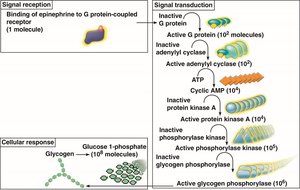
The fight-or-flight response illustrates how a single epinephrine molecule can trigger the breakdown of many glycogen molecules in muscle cells, amplifying the signal. - Signal amplification: One hormone molecule leads to a large cellular response. - Pathway: Epinephrine binds to a receptor, activating relay molecules and enzymes that break down glycogen into glucose. 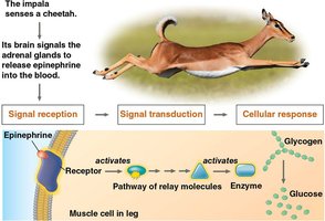
G-Protein Coupled Receptor (GPCR) Signaling
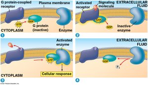
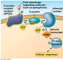
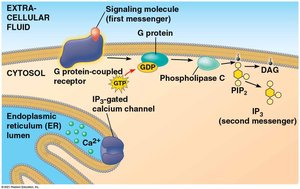
GPCRs are a major class of cell-surface receptors involved in many signaling pathways. - Reception: Ligand binds to GPCR, causing a conformational change. - Transduction: GPCR activates a G protein, which then activates an enzyme (e.g., adenylyl cyclase). - Second messengers: Adenylyl cyclase converts ATP to cAMP, which activates protein kinase A (PKA) and triggers cellular responses. 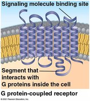
Second Messengers
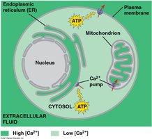
Second messengers are small, water-soluble molecules or ions that rapidly diffuse inside cells, amplifying the signal. - cAMP: Produced from ATP by adenylyl cyclase; activates downstream responses. - Other examples: Calcium ions (Ca2+), inositol triphosphate (IP3). 
Cellular Responses and Pathway Integration
Signal Response Depends on Cell Type
The same signal can produce different responses in different cell types, depending on receptor expression and pathway branching. - Pathway branching: A single signal can activate multiple responses. - Cross-talk: Pathways can interact, integrating multiple signals. 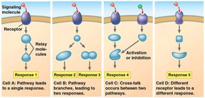
Types of Cellular Responses
Cellular responses to signals can be rapid or slow: - Rapid: Modification of existing proteins (e.g., phosphorylation). - Slow: Changes in gene expression, leading to altered protein synthesis.
Receptor Tyrosine Kinases (RTKs)
RTK Signaling in Cell Division
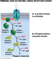
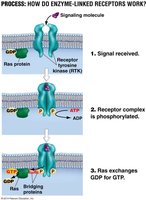
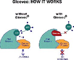
RTKs are enzyme-linked receptors that play a key role in cell growth and division. - Reception: Ligand binding causes dimerization and autophosphorylation of tyrosines. - Transduction: Adaptor proteins link RTK to Ras, triggering a phosphorylation cascade. - Response: Amplified signal drives cell division. - Dysregulation: Overactive RTKs can lead to uncontrolled cell division and cancer. 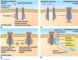
Other Signaling Mechanisms
Ligand-Gated Ion Channels
Ligand-gated ion channels convert chemical signals into electrical responses by allowing ions to flow across the membrane. - Example: Neuron-to-muscle signaling for contraction (Na+ / Ca2+ influx). 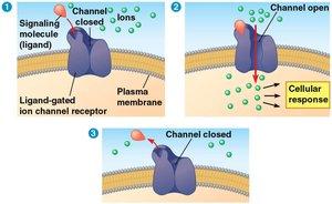
Intracellular Receptors
Intracellular receptors are located in the cytoplasm or nucleus and bind small or hydrophobic hormones. - Hormone–receptor complex: Acts as a transcription factor, changing gene expression. - Example: Aldosterone regulates blood pressure. 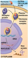
Apoptosis: Programmed Cell Death
Mechanism of Apoptosis
Apoptosis is a controlled process that removes damaged, infected, or aged cells. - Reception: Cell senses external death signals or internal damage. - Transduction: Loss of survival signals activates caspases. - Response: Caspases and nucleases dismantle the cell; contents are packaged for removal. - Termination: Signals turn off; scavenger cells clear apoptotic bodies. 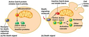
Importance of Apoptosis
Apoptosis is critical for development and tissue homeostasis. - Removes: Infected, damaged, or aged cells. - Prevents: Harmful leakage of enzymes/toxins. - Dysregulation: Too little apoptosis can cause cancer; too much can cause neurodegenerative diseases. 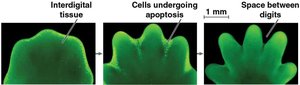
Hormonal Regulation and Homeostasis
Insulin and Glucagon: Activation and Termination
Insulin and glucagon regulate blood glucose levels through precise activation and termination. - Insulin: Activated when blood glucose is high; promotes glucose uptake and storage. - Glucagon: Activated when glucose is low; promotes glucose release from liver. - Termination: Prevents over-correction and maintains homeostasis. 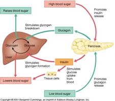
Homeostasis
Homeostasis is the maintenance of stable internal conditions despite external changes, requiring precise hormonal signaling. 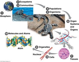
Hormone Diversity
Examples of Hormones
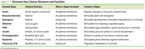
Hormones have diverse structures and functions, affecting both plants and animals.
Hormone Name | Chemical Structure | Where is Signal Received? | Function of Signal |
|---|---|---|---|
Auxin | Small organic compound | At plasma membrane | Signal changes in long axis of plant body |
Brassinosteroids | Steroid | At plasma membrane | Stimulate plant cell elongation |
Estrogens | Steroid | Inside cell | Stimulate development of female characteristics in animals |
Ethylene | C2H4 gas | At plasma membrane | Stimulates fruit ripening, regulates plant growth |
FSH | Glycoprotein | At plasma membrane | Stimulates egg maturation, sperm production in animals |
Insulin | Protein, 51 amino acids | At plasma membrane | Stimulates glucose uptake in animal cells |
Progesterone | Steroid | Inside cell | Stimulates pregnancy functions in animals |
Testosterone | Steroid | Inside cell | Stimulates development of male characteristics in animals |
Thyroxine (T4) | Modified amino acid | Inside cell | Regulates metabolism in animals |