 Indietro
Chapter 12-13: Cell Cycle, Mitosis, and Meiosis
Guida di studio - Note intelligenti
 Appunti personalizzati basati sui tuoi materiali, ampliati con definizioni chiave, esempi e contesto.
The Cell Cycle
Overview of Cell Division
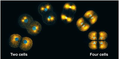
Cell division is a fundamental process that enables organisms to grow, develop, and maintain their tissues. It is essential for reproduction, growth, and repair in multicellular organisms. Most cell division results in genetically identical daughter cells, ensuring continuity of life.
Cell division allows single-celled organisms to reproduce and multicellular organisms to develop, renew, and repair tissues.
Accurate distribution of genetic material is crucial for maintaining genetic integrity across generations.
Cellular Organization of Genetic Material
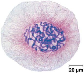
Genetic material in cells is organized into chromosomes, which are composed of DNA and proteins. The complete set of DNA in a cell is called its genome.
Genome: All DNA in a cell; can be a single molecule (prokaryotes) or multiple molecules (eukaryotes).
Chromosomes: DNA molecules packaged with proteins; carry genes.
Chromatin: Complex of DNA and protein in eukaryotic chromosomes; condenses during cell division.
Somatic cells: Nonreproductive cells with two sets of chromosomes.
Gametes: Reproductive cells (sperm and eggs) with half the chromosome number of somatic cells.
Chromosome Duplication and Distribution
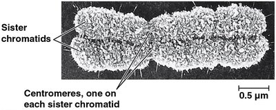
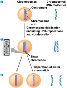
Before cell division, chromosomes are duplicated, resulting in two sister chromatids joined at the centromere. During division, chromatids separate and are distributed to daughter cells.
Sister chromatids: Joined copies of a chromosome after DNA replication.
Centromere: Region where chromatids are most closely attached.
Types of Cell Division
Eukaryotic cell division consists of two main processes: mitosis and cytokinesis. Gametes are produced by meiosis, which results in nonidentical daughter cells with half the chromosome number.
Mitosis: Division of genetic material in the nucleus.
Cytokinesis: Division of the cytoplasm.
Meiosis: Specialized division producing gametes.
Phases of the Cell Cycle
Cell Cycle Structure
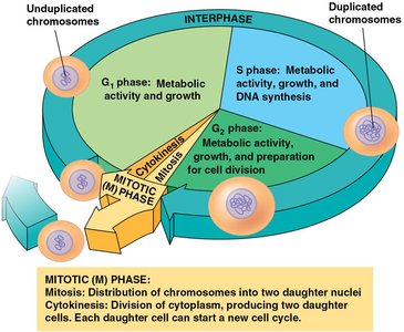
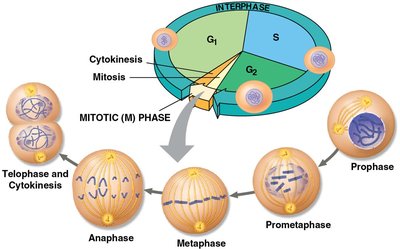
The cell cycle is composed of the mitotic (M) phase and interphase. Interphase is subdivided into G1, S, and G2 phases.
G1 phase: First gap, cell growth.
S phase: Synthesis, DNA replication.
G2 phase: Second gap, preparation for division.
M phase: Mitosis and cytokinesis.
Stages of Mitosis
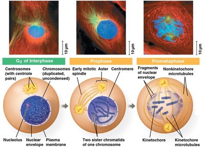
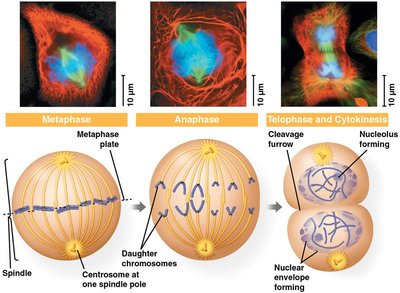
Mitosis is conventionally divided into five stages: prophase, prometaphase, metaphase, anaphase, and telophase.
Prophase: Chromosomes condense, spindle forms.
Prometaphase: Nuclear envelope fragments, spindle microtubules attach to kinetochores.
Metaphase: Chromosomes align at metaphase plate.
Anaphase: Sister chromatids separate and move to opposite poles.
Telophase: Nuclear envelope reforms, chromosomes decondense.
The Mitotic Spindle
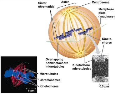
The mitotic spindle is a microtubule-based structure that orchestrates chromosome movement during mitosis. It includes centrosomes, spindle microtubules, and asters.
Centrosome: Microtubule-organizing center; replicates and migrates to cell poles.
Kinetochore: Protein complex at centromere; attaches to spindle microtubules.
Metaphase plate: Imaginary plane where chromosomes align during metaphase.
Cytokinesis
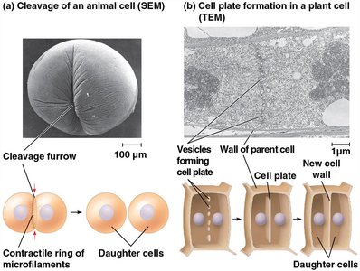
Cytokinesis is the process by which the cytoplasm divides, forming two daughter cells. In animal cells, this occurs via cleavage; in plant cells, a cell plate forms.
Cleavage furrow: Shallow groove in animal cells indicating cytokinesis.
Cell plate: Structure in plant cells that leads to new cell wall formation.
Binary Fission in Prokaryotes
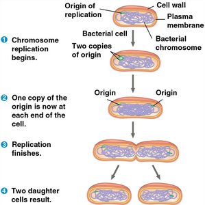
Prokaryotes reproduce by binary fission, a simpler form of cell division. The chromosome replicates, and the cell splits into two.
Origin of replication: Starting point for DNA replication.
Binary fission: Chromosome replication, segregation, and cell division.
Cell Cycle Regulation
Cell Cycle Control System
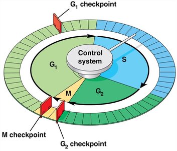
The cell cycle is regulated by a control system with checkpoints. Internal and external signals determine whether the cell proceeds through the cycle.
Checkpoints: G1, G2, and M phase checkpoints monitor cell readiness.
G0 phase: Nondividing state entered if cell does not receive go-ahead signal.
Cyclins and Cyclin-Dependent Kinases (Cdks)
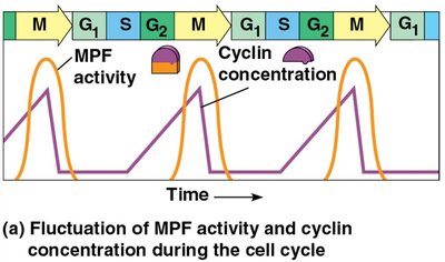
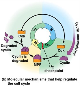
Regulatory proteins called cyclins and Cdks control cell cycle progression. Cyclin concentrations fluctuate, activating Cdks at specific points.
MPF (Maturation-Promoting Factor): Cyclin-Cdk complex that triggers entry into M phase.
MPF activity peaks correspond to cyclin concentration peaks.
Summary of Mitotic (M) Phase
The mitotic phase includes mitosis and cytokinesis, resulting in two genetically identical daughter cells. Each daughter cell can start a new cell cycle.
Meiosis and Sexual Life Cycles
Inheritance and Chromosomes
Offspring inherit genes from parents via chromosomes. Variation arises from the combination of parental genes and genetic mechanisms.
Genes: Units of heredity, segments of DNA.
Gametes: Reproductive cells transmitting genes.
Locus: Specific position of a gene on a chromosome.
Sets of Chromosomes in Human Cells
Human somatic cells have 23 pairs of chromosomes, including autosomes and sex chromosomes. Homologous chromosomes carry genes for the same traits.
Karyotype: Ordered display of chromosome pairs.
Diploid (2n): Two sets of chromosomes; for humans, 2n = 46.
Haploid (n): Single set of chromosomes; for humans, n = 23.
Human Life Cycle
Fertilization unites gametes, forming a zygote. The zygote divides by mitosis to become an adult. Gametes are produced by meiosis, maintaining chromosome number across generations.
Types of Sexual Life Cycles
Animals, plants, and fungi exhibit different sexual life cycles, but all involve alternation between haploid and diploid stages.
Meiosis: Reducing Chromosome Number
Meiosis consists of two consecutive divisions (meiosis I and II), resulting in four haploid daughter cells. Each cell is genetically distinct.
Meiosis I: Homologous chromosomes separate.
Meiosis II: Sister chromatids separate (similar to mitosis).
Comparison of Mitosis and Meiosis
Meiosis differs from mitosis in three key ways: synapsis and crossing over, alignment of homologous pairs, and separation of homologs. Cohesins are cleaved differently in each process.
Genetic Variation and Evolution
Origins of Genetic Variation
Genetic variation arises from mutations, independent assortment, crossing over, and random fertilization. These mechanisms contribute to evolution by creating diverse offspring.
Independent assortment: Chromosomes assort independently into gametes; for humans, more than 8 million combinations.
Crossing over: Exchange of genetic material between homologous chromosomes, producing recombinant chromosomes.
Random fertilization: Any sperm can fuse with any egg, resulting in about 70 trillion possible combinations.
Evolutionary Significance
Genetic variation within populations is essential for natural selection and adaptation. Sexual reproduction and mechanisms like crossing over and independent assortment ensure diversity.
Additional info: These notes expand on brief points from the original slides, providing definitions, examples, and context for General Biology students. All included images directly reinforce the adjacent explanations and are strictly relevant to the content.
