 Indietro
Cell Division, Cancer, and Chemotherapy: Step-by-Step Study Guidance
Guida di studio - Note intelligenti
 Appunti personalizzati basati sui tuoi materiali, ampliati con definizioni chiave, esempi e contesto.
Q1. Why is cell division necessary? Describe three situations in which cell division occurs.
Background
Topic: Cell Division and Its Importance
This question tests your understanding of why cells divide and the biological contexts in which cell division is essential.
Key Terms:
Cell division: The process by which a parent cell divides into two or more daughter cells.
Mitosis: Division of somatic cells for growth and repair.
Meiosis: Division that produces gametes for sexual reproduction.
Step-by-Step Guidance
Think about why organisms need new cells. Consider growth, repair, and reproduction.
Identify three distinct situations where cell division is required (e.g., development from a fertilized egg, healing wounds, producing gametes).
For each situation, briefly explain how cell division contributes to the process.
Try solving on your own before revealing the answer!
Final Answer:
Cell division is necessary for:
Growth: Organisms grow by increasing their cell number through mitosis.
Repair: Damaged tissues are repaired by replacing dead or injured cells.
Reproduction: In sexually reproducing organisms, meiosis produces gametes (sperm and eggs).
These processes ensure survival, maintenance, and propagation of life.
Q2. Describe the organization of the genetic material by defining the following terms: Genome, Chromosomes, Chromatin, Somatic cells, Gametes.
Background
Topic: Genetic Material Organization
This question tests your knowledge of how genetic material is structured and classified in cells.
Key Terms:
Genome: The complete set of genetic material in an organism.
Chromosomes: Structures that carry genetic information, made of DNA and proteins.
Chromatin: The complex of DNA and proteins that forms chromosomes.
Somatic cells: Body cells (non-reproductive), diploid.
Gametes: Reproductive cells (sperm/egg), haploid.
Step-by-Step Guidance
Start by defining the genome as the total genetic content.
Explain how chromosomes are organized and what they contain.
Describe chromatin and its role in packaging DNA.
Distinguish between somatic cells and gametes in terms of chromosome number.
Try solving on your own before revealing the answer!
Final Answer:
Genome: All the DNA in a cell, including genes and non-coding regions.
Chromosomes: Linear or circular DNA molecules with associated proteins, visible during cell division.
Chromatin: DNA-protein complex; condenses to form chromosomes during mitosis.
Somatic cells: Diploid cells (2 sets of chromosomes), make up most of the body.
Gametes: Haploid cells (1 set of chromosomes), produced by meiosis for sexual reproduction.
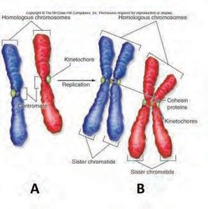
Q3. Use the figures to describe: Homologous Chromosomes and Sister Chromatids.
Background
Topic: Chromosome Structure
This question tests your ability to distinguish between homologous chromosomes and sister chromatids using visual aids.
Key Terms:
Homologous chromosomes: Pair of chromosomes, one from each parent, similar in shape and gene content.
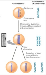
Sister chromatids: Identical copies of a chromosome, joined at the centromere after DNA replication.
Step-by-Step Guidance
Examine the provided figure and identify the homologous chromosomes (pairs of similar chromosomes, one red and one blue).
Locate the sister chromatids (identical copies joined together after replication).
Describe the relationship between homologous chromosomes and sister chromatids in terms of genetic content and structure.
Try solving on your own before revealing the answer!
Final Answer:
Homologous chromosomes are pairs (one from each parent) that carry the same genes but may have different alleles. Sister chromatids are identical copies of a single chromosome, joined at the centromere, formed after DNA replication. During cell division, sister chromatids separate to become individual chromosomes.
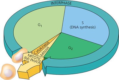
Q4. Describe the overall process of the cell cycle. Describe the general function of each phase. Which is the longest part?
Background
Topic: Cell Cycle Phases
This question tests your understanding of the cell cycle and the function of each phase.
Key Terms and Formula:
Cell cycle: The series of events that cells go through as they grow and divide.
Phases: G1 (growth), S (DNA synthesis), G2 (preparation for mitosis), M (mitosis and cytokinesis).
Step-by-Step Guidance
List the main phases of the cell cycle: G1, S, G2, and M.
Describe the function of each phase (e.g., G1: cell growth, S: DNA replication, G2: preparation for division, M: mitosis/cytokinesis).
Identify which phase is typically the longest and explain why.
Try solving on your own before revealing the answer!
Final Answer:
The cell cycle consists of G1 (growth), S (DNA synthesis), G2 (preparation), and M (mitosis/cytokinesis). Interphase (G1, S, G2) is the longest part, as cells spend most of their time growing and preparing for division.
Q5. Cell division occurs in multiple steps. Describe major features of each step below: Interphase (G1, S phase, G2), Mitosis (Prophase, Metaphase, Anaphase, Telophase), Cytokinesis.
Background
Topic: Steps of Cell Division
This question tests your knowledge of the sequence and features of cell division.
Key Terms:
Interphase: G1 (growth), S (DNA replication), G2 (preparation).
Mitosis: Prophase (chromosome condensation), Metaphase (alignment), Anaphase (separation), Telophase (reformation).
Cytokinesis: Division of cytoplasm.
Step-by-Step Guidance
Describe what happens in each sub-phase of interphase (G1, S, G2).
Explain the sequence of mitosis steps and their main features.
Summarize the process of cytokinesis and its importance.
Try solving on your own before revealing the answer!
Final Answer:
G1: Cell grows and prepares for DNA replication.
S: DNA is replicated.
G2: Cell prepares for mitosis.
Prophase: Chromosomes condense, spindle forms.
Metaphase: Chromosomes align at the cell equator.
Anaphase: Sister chromatids separate.
Telophase: Nuclear envelope reforms.
Cytokinesis: Cytoplasm divides, forming two cells.
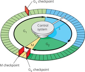
Q6. Cells can continue to grow out of control when checkpoint processes fail. Describe the checkpoints that regulate the cell cycle.
Background
Topic: Cell Cycle Checkpoints
This question tests your understanding of cell cycle regulation and the importance of checkpoints.
Key Terms:
Checkpoints: Control mechanisms that ensure proper cell cycle progression.
G1, G2, M checkpoints: Specific points where the cell assesses readiness to proceed.
Step-by-Step Guidance
Identify the main checkpoints: G1, G2, and M.
Describe what each checkpoint checks for (e.g., DNA damage, proper chromosome alignment).
Explain what happens if a checkpoint fails.
Try solving on your own before revealing the answer!
Final Answer:
G1 checkpoint: Checks for DNA damage and cell size.
G2 checkpoint: Ensures DNA is fully replicated and undamaged.
M checkpoint: Verifies chromosome alignment and spindle attachment.
If checkpoints fail, cells may divide uncontrollably, leading to cancer.
Q7. Cancer results from out of control cell growth. There are two classes of genes that can result in cancer. Describe the difference between each and how they cause cancer.
Background
Topic: Cancer Genetics
This question tests your understanding of oncogenes and tumor suppressor genes.
Key Terms:
Oncogenes: Genes that promote cell division; mutations can lead to uncontrolled growth.
Tumor suppressor genes: Genes that inhibit cell division; loss of function can lead to cancer.
Step-by-Step Guidance
Define oncogenes and tumor suppressor genes.
Explain how mutations in each class contribute to cancer.
Compare their roles in normal and cancerous cells.
Try solving on your own before revealing the answer!
Final Answer:
Oncogenes are mutated forms of genes that normally stimulate cell division; when overactive, they cause excessive growth. Tumor suppressor genes normally inhibit cell division; when inactivated, cells divide uncontrollably. Both types of mutations can lead to cancer.
Q8. How many copies need to be mutated in a tumor suppressor gene to cause cancer? In a proto-oncogene?
Background
Topic: Gene Mutation and Cancer
This question tests your understanding of the genetic requirements for cancer development.
Key Terms:
Tumor suppressor gene: Requires both copies to be mutated for loss of function.
Proto-oncogene: Only one copy needs to be mutated to become an oncogene.
Step-by-Step Guidance
Recall the difference between dominant and recessive mutations.
Apply this to tumor suppressor genes (recessive) and proto-oncogenes (dominant).
State how many alleles must be mutated for each gene type to contribute to cancer.
Try solving on your own before revealing the answer!
Final Answer:
Tumor suppressor genes require both copies (alleles) to be mutated for cancer to develop. Proto-oncogenes only need one copy mutated to become an oncogene and promote cancer.
Q9. Describe the role of ras in the cell. How does a malfunctional ras result in cancer?
Background
Topic: Ras Protein and Cell Signaling
This question tests your understanding of the ras protein's function and its impact on cell division.
Key Terms:
Ras: A GTPase involved in transmitting signals for cell growth.
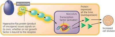
Mutation: Can cause ras to be permanently active, leading to uncontrolled cell division.
Step-by-Step Guidance
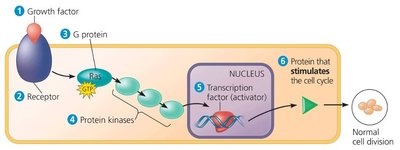
Describe the normal role of ras in cell signaling pathways.
Explain how ras activation leads to cell division.
Discuss what happens when ras is mutated and cannot be inactivated.
Try solving on your own before revealing the answer!
Final Answer:
Ras normally transmits growth signals from the cell surface to the nucleus, promoting cell division. A malfunctional (mutated) ras is always active, causing continuous cell division and contributing to cancer.
Q10. Describe how the role of ras in the cell. How does a malfunctional ras result in cancer?
Background
Topic: Ras Protein and Cancer
This question is similar to Q9 and reinforces your understanding of ras mutations.
Key Terms:
Ras: Signal transduction protein.
Oncogene: Mutated ras acts as an oncogene.
Step-by-Step Guidance
Review the normal function of ras in cell division.
Explain how a mutation in ras leads to constant signaling.
Connect this to the development of cancer.
Try solving on your own before revealing the answer!
Final Answer:
Mutated ras is always "on," causing cells to divide uncontrollably, which is a hallmark of cancer.
Q11. Describe estrogen, progesterone, and HER2 receptor signaling in the development of breast cancer.
Background
Topic: Hormone and Growth Factor Signaling in Cancer
This question tests your understanding of how hormone and growth factor receptors contribute to breast cancer.
Key Terms:
Estrogen/progesterone receptors: Hormone receptors that stimulate cell growth.
HER2: Growth factor receptor; overexpression leads to increased cell division.
Step-by-Step Guidance
Describe how estrogen and progesterone receptors signal cell growth in breast tissue.
Explain the role of HER2 in cell division and how its overexpression affects cancer development.
Connect receptor signaling to uncontrolled cell proliferation in breast cancer.

Try solving on your own before revealing the answer!
Final Answer:
Estrogen and progesterone receptors promote cell growth when activated. HER2 is a growth factor receptor; its overexpression leads to excessive cell division. These signaling pathways drive breast cancer development when dysregulated.
Q12. How does Herceptin work as a therapeutic?
Background
Topic: Targeted Cancer Therapy
This question tests your understanding of how Herceptin targets HER2-positive breast cancer.
Key Terms:
Herceptin: Monoclonal antibody therapy.
HER2: Target receptor for Herceptin.
Step-by-Step Guidance
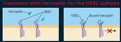
Describe the mechanism of Herceptin binding to HER2 receptors.
Explain how this blocks HER2 signaling and inhibits cell division.
Connect this to reduced tumor growth in HER2-positive breast cancer.
Try solving on your own before revealing the answer!
Final Answer:
Herceptin binds to HER2 receptors, blocking their activity and preventing excessive cell division. This slows or stops the growth of HER2-positive breast cancer cells.
