 Indietro
Cell Division, Chromosomes, and Inheritance: Study Guide
Guida di studio - Note intelligenti
 Appunti personalizzati basati sui tuoi materiali, ampliati con definizioni chiave, esempi e contesto.
Cell Division: Types and Importance
Introduction to Cell Division
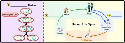
Cell division is a fundamental biological process by which a single parent cell divides to produce two or more daughter cells. This process is essential for reproduction, growth, and tissue repair in all living organisms.
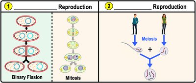
Binary Fission: A type of cell division in prokaryotes (bacteria and archaea) where a single cell splits into two identical cells.
Mitosis: Eukaryotic cell division that produces genetically identical somatic (body) cells.
Meiosis: Eukaryotic cell division that produces genetically diverse gametes (sex cells).
Human somatic cells: Diploid (2n), containing two copies of each chromosome.
Human gametes: Haploid (n), containing one copy of each chromosome.
Importance of Cell Division
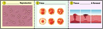
Cell division is crucial for reproduction, fetal development, and tissue repair. Before division, cells must replicate their DNA to ensure each daughter cell receives a complete set of genetic material.
Reproduction: Produces new organisms.
Fetal Development: Enables growth from a single cell to a multicellular organism.
Tissue Repair: Replaces damaged or dead cells.
Organization of DNA in the Cell
Genome and Chromatin Structure
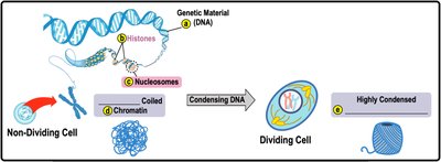
The genome is the complete set of DNA in a cell. DNA associates with proteins called histones to form nucleosomes, which further coil to create chromatin or chromosomes depending on the cell's state.
Genetic Material: DNA molecules that determine inherited traits.
Histones: Proteins around which DNA wraps to form nucleosomes.
Nucleosomes: Units of eight histone proteins with DNA wrapped around them.
Chromatin: Loosely packed nucleosomes in non-dividing cells.
Chromosomes: Highly condensed nucleosomes in dividing cells.
DNA Replication and Chromosome Structure
DNA Replication
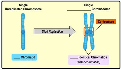
Before cell division, DNA is replicated to produce identical copies. Each replicated chromosome consists of two sister chromatids joined at the centromere.
Chromatid: One of two identical halves of a replicated chromosome.
Centromere: The region where sister chromatids are attached.
The Cell Cycle
Phases of the Cell Cycle
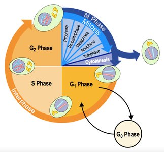
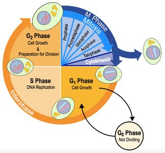
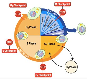
The cell cycle describes the sequence of events from cell formation to division. It consists of interphase (cell growth and DNA replication) and the M phase (mitosis and cytokinesis).
Interphase: Includes G1 (growth), S (DNA synthesis), G2 (preparation for division), and G0 (non-dividing).
M Phase: Includes mitosis (nuclear division) and cytokinesis (cytoplasmic division).
Centrosomes and Mitotic Spindle
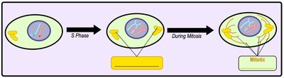
During S phase, centrosomes are replicated. They organize the mitotic spindle, which separates chromosomes during mitosis.
Centrosome: Microtubule-organizing center.
Mitotic Spindle: Structure that moves chromosomes during cell division.
Phases of Mitosis
Overview of Mitosis
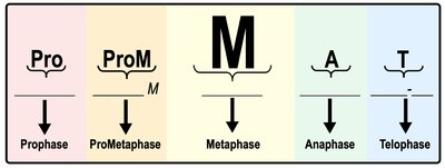
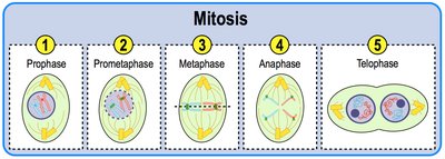
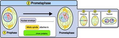
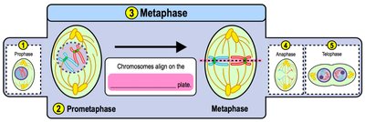
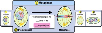
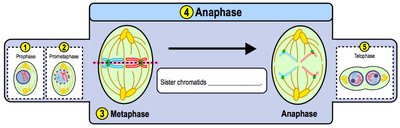
Mitosis is the process by which a diploid cell divides to produce two genetically identical daughter cells. It consists of five phases: prophase, prometaphase, metaphase, anaphase, and telophase.
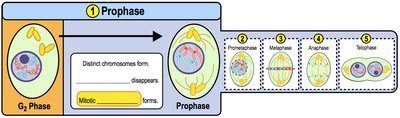
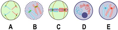
Prophase: Chromatin condenses, nucleolus disappears, centrosomes move to poles.
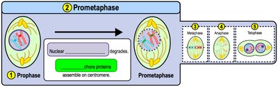
Prometaphase: Nuclear envelope breaks down, spindle attaches to chromosomes.
Metaphase: Chromosomes align at the metaphase plate.
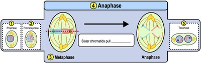
Anaphase: Sister chromatids are pulled apart to opposite poles.
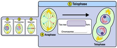
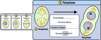
Telophase: Chromosomes decondense, nuclear envelope reforms, two nuclei form.
Cytokinesis
Animal Cell Cytokinesis
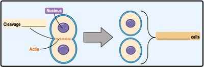
After mitosis, cytokinesis divides the cytoplasm, resulting in two identical daughter cells. In animal cells, a cleavage furrow forms through the action of actin and myosin.
Cleavage Furrow: Indentation at the cell center where division occurs.
Plant Cell Cytokinesis
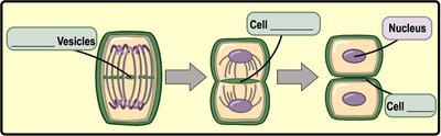
Plant cells form a cell plate from Golgi-derived vesicles, which develops into a new cell wall separating the daughter cells.
Cell Plate: Structure that becomes the cell wall in plant cells.
Cell Cycle Regulation
Checkpoints and Regulation
The cell cycle is regulated by checkpoints that ensure proper DNA replication and division. Growth factors stimulate division, while proteins like p53 can trigger repair or apoptosis if errors are detected.
Checkpoints: G1, S, G2, and M checkpoints control cell cycle progression.
Apoptosis: Programmed cell death if errors cannot be repaired.
Genes, Alleles, and Chromosome Structure
Genes and Alleles
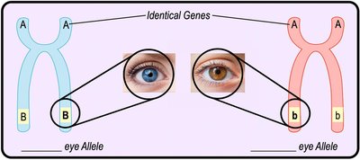
Genes are segments of DNA encoding proteins that determine traits. Alleles are different versions of a gene, often represented by capital and lowercase letters.
Example: B = blue eyes, b = brown eyes.
Haploid vs. Diploid Cells
Cell ploidy refers to the number of sets of chromosomes in a cell. Haploid cells (n) have one set, while diploid cells (2n) have two sets, one from each parent.
Example: Human somatic cells are diploid; gametes are haploid.
Homologous Chromosomes
Homologous chromosomes are pairs that are similar in size and shape and carry the same genes, but may have different alleles.
Karyotype: Ordered display of all chromosomes in a cell.
Autosomes: Non-sex chromosomes.
Sex Chromosomes: X and Y chromosomes determine sex.
Meiosis and Genetic Variation
Meiosis: Process and Significance
Meiosis is a form of cell division that produces haploid gametes, enabling sexual reproduction and genetic diversity. It consists of two rounds: Meiosis I (separates homologous chromosomes) and Meiosis II (separates sister chromatids).
Crossing Over: Exchange of genetic material between homologous chromosomes during Prophase I.
Independent Assortment: Random alignment of homologous pairs during Metaphase I.
Nondisjunction: Failure of chromosomes to separate, leading to aneuploid cells.
Inheritance Patterns and Mendelian Genetics
Mendel’s Experiments and Laws
Gregor Mendel discovered the principles of inheritance using pea plants. He formulated the Law of Segregation and the Law of Independent Assortment.
Law of Segregation: Alleles of a gene separate during gamete formation.
Law of Independent Assortment: Alleles of different genes assort independently during meiosis.
Punnett Squares and Genetic Probability
Punnett squares are diagrams used to predict the genotypes and phenotypes of offspring from genetic crosses. Probability rules (multiplication and addition) are used to calculate outcomes.
Non-Mendelian Genetics
Some traits do not follow Mendel’s laws, including incomplete dominance, codominance, polygenic inheritance, pleiotropy, and multifactorial characters.
Incomplete Dominance: Heterozygotes show intermediate phenotypes.
Codominance: Both alleles are fully expressed.
Polygenic Inheritance: Multiple genes affect a single trait.
Pleiotropy: One gene affects multiple traits.
Multifactorial Characters: Traits influenced by genetic and environmental factors.
Pedigrees and Patterns of Inheritance
Pedigree Analysis
Pedigrees are charts used to track inheritance patterns across generations. Autosomal and sex-linked inheritance can be identified using pedigree analysis.
Autosomal Dominant: Disorder appears in every generation.
Autosomal Recessive: Disorder may skip generations.
Sex-Linked: Disorders often affect males more than females.
X-Inactivation
Mechanism and Consequences
In female mammals, one X chromosome is randomly inactivated in each cell, forming a Barr body. This prevents double expression of X-linked genes and can result in mosaic phenotypes.
Barr Body: Inactive X chromosome in female cells.
