 Indietro
Cell Division: Mitosis, Meiosis, and Chromosomal Inheritance
Guida di studio - Note intelligenti
 Appunti personalizzati basati sui tuoi materiali, ampliati con definizioni chiave, esempi e contesto.
Mitosis: Basic Cell Division
Purpose and Cell Types
Mitosis is a fundamental process for growth and repair in multicellular organisms. It occurs primarily in somatic (body) cells of animals and in both sporophyte (2n) and gametophyte (1n) cells of plants.
Purpose: Growth and repair (healing).
Cell Types: Somatic cells in animals; sporophyte and gametophyte cells in plants.
Ploidy: One diploid (2n) cell divides once to produce two genetically identical diploid (2n) cells.
The Cell Cycle
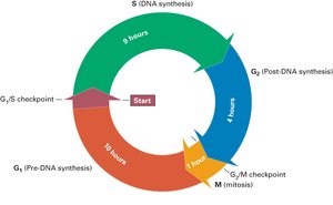
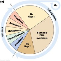
The cell cycle consists of interphase (G1, S, G2) and mitosis (M phase). Checkpoints regulate transitions between phases to ensure proper cell division.
Interphase: G1 (cell growth), S (DNA synthesis), G2 (preparation for mitosis).
Mitosis: Division of the nucleus and cytoplasm.
Checkpoints: G1/S and G2/M checkpoints control progression.
Mitosis Overview
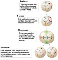
During mitosis, chromosome number remains unchanged, and two daughter cells are produced. Chromosome behavior is illustrated in organisms with homologous chromosome pairs.
Chromosome Number: Maintained throughout mitosis.
Daughter Cells: Two genetically identical cells.
The Phases of Mitosis
Interphase
Cell growth and DNA replication occur. Chromosomes are not yet condensed.
Prophase
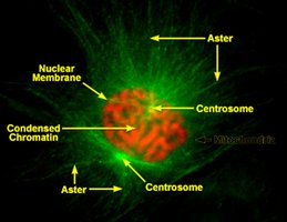
Centrioles duplicate (microtubule organizing centers).
Nuclear membrane breaks down.
Microtubule spindle forms.
Chromosomes condense; sister chromatids attach at the centromere.
Homologous chromosomes do not pair.
Prometaphase and Metaphase
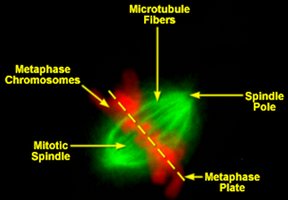
Chromosomes move to the metaphase plate (prometaphase).
Chromosomes align at the metaphase plate (metaphase).
Anaphase
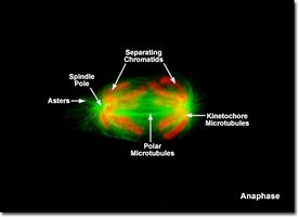
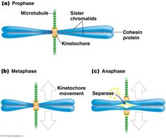
Sister chromatids separate due to the enzyme separase cleaving cohesin.
Chromatids move to opposite poles (chromosome disjunction).
Errors in this process are called non-disjunction.
Telophase and Cytokinesis
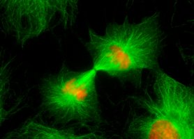
Chromosomes reach the poles.
Chromosomes decondense.
Nuclear envelope reforms.
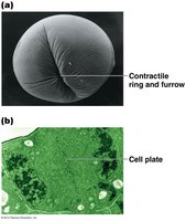
Cytokinesis divides the cytoplasm, forming two daughter cells.
Meiosis: Making Gametes
Purpose and Cell Types
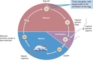
Meiosis is essential for sexual reproduction, producing gametes (sperm and eggs) that are genetically distinct.
Purpose: Reproduction.
Cell Types: Germline cells (gonads).
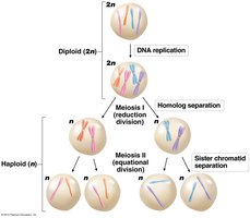
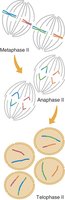
Ploidy: One diploid (2n) cell divides twice to produce four haploid (1n) cells.
Meiosis: Cell Division in Two Parts
Meiosis consists of two sequential divisions: Meiosis I (reductional) and Meiosis II (equational).
Mitosis vs. Meiosis
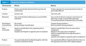
Mitosis and meiosis differ in purpose, mechanism, and outcome. Mitosis produces identical cells, while meiosis produces genetically diverse gametes.
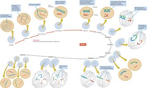
Meiosis I: Hallmark Events
Homologous Chromosome Pairing: Homologs pair up.
Crossing Over: Exchange of genetic material between homologs.
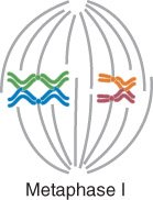
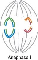
Segregation: Homologs separate, reducing chromosome number to haploid.
Sister chromatids remain together.
Prophase I Substages
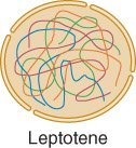
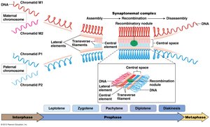
Leptotene: Chromosome condensation begins; chromomeres visible.
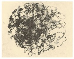
Zygotene: Homologous chromosomes pair (synapsis).
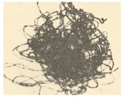
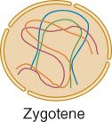
Pachytene: Synapsis completed; crossing over occurs.
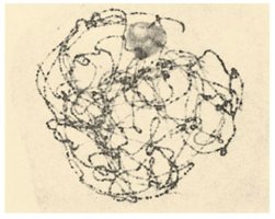
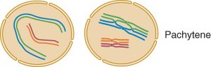
Diplotene: Synaptonemal complex breaks down; chiasmata (crossing over sites) visible.
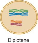
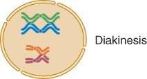
Diakinesis: Homologs repel each other but remain attached at chiasmata.
The Synaptonemal Complex
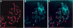
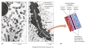
The synaptonemal complex is a protein structure that forms between homologous chromosomes during prophase I, facilitating crossing over and recombination.
Independent Assortment
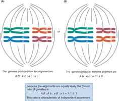
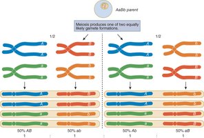
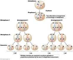
Independent assortment refers to the random alignment and separation of homologous chromosomes during meiosis I, resulting in genetic variation.
For genotype AaBb, four gamete combinations (AB, ab, Ab, aB) occur with equal likelihood.
Physical basis: Random alignment of nonhomologous chromosomes at metaphase I.
Chromosomal Basis of Sex Determination
XX-XY System
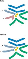
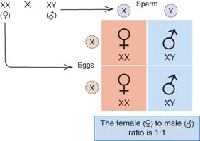
Sex determination in mammals and many insects is based on the presence of X and Y chromosomes. Females are homogametic (XX), while males are heterogametic (XY).
Females: XX
Males: XY
X-linked Inheritance
Experimental Demonstration
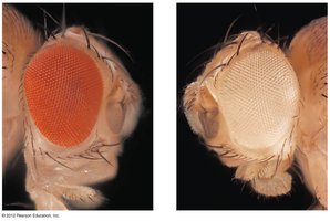
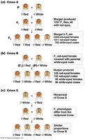
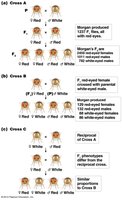
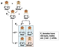
The white mutation in Drosophila was the first experimental proof that genes are located on chromosomes. Reciprocal crosses for the X-linked eye color gene demonstrate inheritance patterns.
Inheritance of X-linked Hemophilia
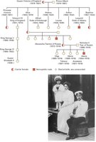
Hemophilia A is an X-linked disorder, and its inheritance can be traced through royal pedigrees, such as the descendants of Queen Victoria.
Summary Table: Comparison of Mitosis and Meiosis
Characteristic | Mitosis | Meiosis |
|---|---|---|
Purpose | Growth and repair; produces identical cells | Sexual reproduction; produces gametes |
Location | Somatic cells | Germline cells |
Mechanism | One division after DNA replication | Two divisions (meiosis I and II) |
Homologous Chromosomes | Do not pair | Pair and recombine during prophase I |
Sister Chromatids | Separate at anaphase | Stay together in meiosis I, separate in meiosis II |
Product | Two identical diploid cells | Four genetically distinct haploid cells |
