 Indietro
Cell Membrane Structure and Function: Study Notes (General Biology)
Guida di studio - Note intelligenti
 Appunti personalizzati basati sui tuoi materiali, ampliati con definizioni chiave, esempi e contesto.
Cell Membranes
Plasma Membrane: Structure and Function
The plasma membrane defines the outer boundary of the cell and is essential for maintaining cellular integrity. It regulates the movement of substances into and out of the cell, acting as a selectively permeable or semi-permeable barrier.
Selective permeability: Allows certain molecules to pass while restricting others.
Boundary function: Separates the internal environment from the external environment.
Fluid-Mosaic Model
The fluid-mosaic model describes the structure of cell membranes. The membrane is dynamic, with components moving laterally within the bilayer, and is composed of many individual pieces.
Fluid: Components (lipids and proteins) move freely, mostly laterally.
Mosaic: The membrane is made up of various molecules, including phospholipids, cholesterol, glycolipids, and proteins.
Major Components of the Cell Membrane
Phospholipids
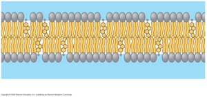
Cholesterol
Glycolipids
Proteins

Phospholipids
Phospholipids are amphipathic molecules, meaning they have both hydrophilic (water-loving) and hydrophobic (water-fearing) regions. They form a phospholipid bilayer with hydrophobic tails hidden inside and hydrophilic heads exposed to the aqueous environment.
Bilayer structure: Two layers of phospholipids.
Hydrophobic interior: Prevents passage of polar molecules.
Hydrophilic exterior: Interacts with water.
Lateral movement: Phospholipids can move within the bilayer.
Cholesterol
Cholesterol is a steroid present in animal cell membranes. It plays a crucial role in modulating membrane fluidity.
Restricts lateral movement: Stabilizes membrane at higher temperatures.
Increases mobility at low temperatures: Acts as an "anti-freeze" to prevent the membrane from becoming too rigid.
Membrane Proteins
Membrane proteins are essential for various cellular functions and are classified based on their association with the membrane.
Integral proteins: Embedded within the membrane, often spanning the bilayer (transmembrane).
Peripheral proteins: Loosely bound to the membrane surface, often attached to integral proteins.

Integral Proteins
Transmembrane proteins: Span the entire membrane.
Hydrophobic regions: Align with the hydrophobic core of the membrane.
Functions: Include transport, enzymatic activity, and signal transduction.
Peripheral Proteins
Surface association: Attached to the membrane surface or to integral proteins.
Functions: Include cell signaling, structural support, and cell-cell interactions.
Functions of Membrane Proteins
Membrane proteins perform a variety of functions essential for cell survival and communication.
Transport: Move substances across the membrane (channels, carriers).
Enzymatic activity: Catalyze reactions at the membrane surface.
Signal transduction: Relay signals from outside to inside the cell.
Cell-cell recognition: Allow cells to identify each other (often via glycoproteins).
Intercellular joining: Connect cells together (junctions).
Attachment: Anchor the membrane to the cytoskeleton and extracellular matrix (ECM).


Synthesis and Assembly of Cell Membranes
Membrane Asymmetry
Cell membranes are asymmetrical, meaning their components (proteins, lipids, carbohydrates) are not equally distributed on either side. This asymmetry is established during membrane synthesis in the endoplasmic reticulum (ER) and Golgi apparatus.
Distinct faces: Membranes have unique inside and outside surfaces.
Component arrangement: Determined during membrane assembly.
Protein Association with Membranes
Proteins associate with membranes in three main ways:
Transmembrane proteins: Embedded within the membrane.
Peripheral proteins: Attached to the membrane surface.
Secreted proteins: Located in the lumen of vesicles.
Membrane Synthesis Pathway
Membrane synthesis involves the ER and Golgi apparatus, with vesicles transporting membrane components. The relationship of proteins to the membrane is maintained throughout this process.
Vesicle formation: Membrane can pinch off to form vesicles.
Fusion with Golgi: Vesicles fuse with Golgi, maintaining protein orientation.
Modification: Proteins are modified in the Golgi, and membranes bud off.
Final destination: Vesicles may remain in the cell (lysosomes, peroxisomes) or fuse with the plasma membrane, adding new membrane.


Summary Table: Types of Membrane Proteins and Their Association
Type | Location | Function |
|---|---|---|
Transmembrane | Embedded in membrane | Transport, signaling |
Peripheral | Surface of membrane | Support, signaling |
Secreted | Lumen of vesicle | Enzymatic, signaling |
Key Terms and Concepts
Phospholipid bilayer: Fundamental structure of cell membranes.
Amphipathic: Molecule with both hydrophilic and hydrophobic regions.
Fluid-mosaic model: Describes membrane structure and dynamics.
Selective permeability: Ability to regulate passage of substances.
Membrane protein functions: Transport, enzymatic activity, signal transduction, recognition, joining, attachment.
Membrane synthesis: Process involving ER, Golgi, and vesicles.
Equations and Academic Context
While cell membrane structure is not typically described by equations, the permeability of the membrane can be modeled mathematically:
Permeability coefficient (P): Where: D = diffusion coefficient K = partition coefficient d = thickness of membrane
Additional info: The notes above expand on the original content by providing definitions, examples, and academic context for each topic. The summary table and equation are inferred to enhance understanding and exam preparation.
