 Indietro
Cell Signaling and Communication: Mechanisms and Examples
Guida di studio - Note intelligenti
 Appunti personalizzati basati sui tuoi materiali, ampliati con definizioni chiave, esempi e contesto.
Cell Communication and Signaling
Overview of Cell Signaling
Cell signaling is a fundamental process by which cells receive, interpret, and respond to external and internal signals. This process enables multicellular organisms to coordinate activities, adapt to environmental changes, and maintain homeostasis. - Signal reception: Cells detect signaling molecules (ligands) from their environment. - Signal transduction: The signal is relayed and amplified inside the cell through a series of molecular events. - Cellular response: The cell executes a specific response, such as gene expression, enzyme activation, or metabolic changes. 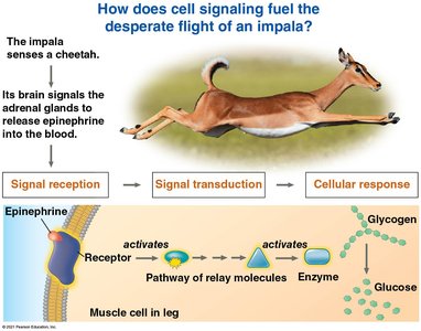
Example: Epinephrine and the Impala's Flight Response
When an impala senses a predator, its brain signals the adrenal glands to release epinephrine (adrenaline) into the bloodstream. This hormone triggers a cascade of events in muscle cells, leading to rapid breakdown of glycogen and release of glucose for energy. - Reception: Epinephrine binds to a receptor on the muscle cell membrane. - Transduction: Relay molecules and enzymes are activated, amplifying the signal. - Response: Glycogen is broken down into glucose, fueling muscle contraction and escape. 
Types of Cell Signaling
Local Signaling: Direct Contact
Cells can communicate directly through physical contact. - Cell junctions: Gap junctions (animals) and plasmodesmata (plants) allow molecules and ions to pass directly between cells. - Cell–cell recognition: Surface molecules on adjacent cells interact, important in development and immune responses. 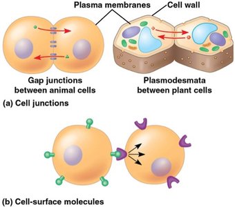
Local Signaling: Secreted Messengers
Cells can also communicate by releasing signaling molecules that affect nearby cells. - Paracrine signaling: Local regulators (e.g., growth factors) are secreted and act on nearby target cells. - Synaptic signaling: Neurons release neurotransmitters across synapses to target cells, enabling rapid communication. 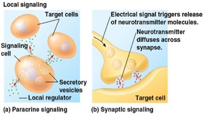
Long-Distance Signaling: Hormones
Hormones are used for communication over long distances. - Endocrine signaling: Hormones are released into the bloodstream and act on distant target cells. - Specificity: Only cells with the appropriate receptor respond to the hormone.
Classes of Chemical Messengers
Peptide/Protein, Steroid, and Amine Messengers
Chemical messengers are classified by their structure and solubility, which determine their transport and receptor location.
Class | Examples | Structure | Solubility | Transport | Receptor Location |
|---|---|---|---|---|---|
Peptide/Protein | Insulin, glucagon, oxytocin | Chain of amino acids | Water-soluble | Dissolved in plasma | Plasma membrane |
Steroid | Aldosterone, cortisol, estrogen | Lipid, four fused rings | Lipid-soluble | Bound to carrier proteins | Intracellular |
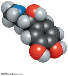
Amine | Epinephrine, norepinephrine, thyroid hormone | Modified amino acid | Epinephrine: water-soluble; Thyroid hormone: lipid-soluble | Epinephrine: free in plasma; Thyroid hormone: on carrier proteins | Epinephrine: membrane; Thyroid hormone: nuclear |
The Three Stages of Cell Signaling
Reception, Transduction, Response, and Reset
Cell signaling involves a series of steps: - Reception: A signaling molecule binds to a receptor protein. - Transduction: The receptor changes shape and initiates a signal transduction pathway. - Response: The cell produces a specific response. - Reset: Components are switched off to allow new signals. 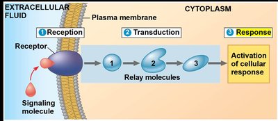
Signal Reception: Types of Receptors
Major Receptor Types
- GPCRs (G protein-coupled receptors): Bind hormones and neurotransmitters. - RTKs (Receptor tyrosine kinases): Bind growth factors and insulin. - Ion channel receptors: Ligand-gated channels for neurotransmitters. - Intracellular receptors: Located in cytoplasm or nucleus; bind steroid and thyroid hormones. 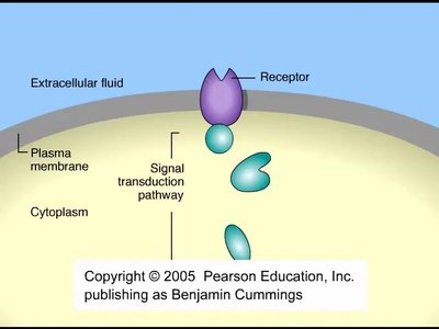
G Protein-Coupled Receptors (GPCRs)
GPCRs are the largest family of cell-surface receptors. - Structure: Transmembrane proteins with a binding site for signaling molecules and an interaction site for G proteins. - Function: Relay signals from outside to inside the cell via G proteins. 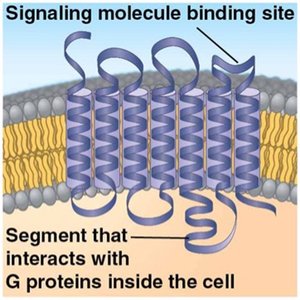
G Proteins: Molecular On/Off Switch
G proteins cycle between active and inactive states. - Inactive: G protein binds GDP. - Activation: Signal binds GPCR, GTP replaces GDP, G protein activates an enzyme. - Reset: G protein hydrolyzes GTP to GDP, returns to inactive state. 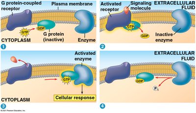
Receptor Tyrosine Kinases (RTKs)
RTKs catalyze the transfer of phosphate groups from ATP to proteins. - Ligands: Growth factors. - Function: Trigger multiple signal transduction pathways. - Clinical relevance: Abnormal RTK function is linked to cancers. 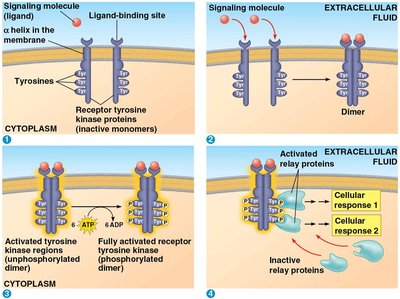
Ligand-Gated Ion Channel Receptors
These receptors act as gates for ions. - Activation: Ligand binding opens the channel, allowing ions to flow. - Reset: Channel closes when ligand leaves. 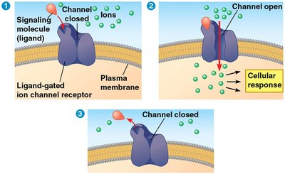
Intracellular Receptors
Located in the cytoplasm or nucleus, these receptors bind hydrophobic messengers. - Example: Steroid hormones like aldosterone cross the membrane and bind intracellular receptors, acting as transcription factors. 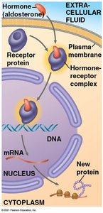
Signal Transduction Pathways
Phosphorylation Cascades
Signal transduction often involves cascades of protein kinases. - Protein kinases: Add phosphate groups to proteins, activating them. - Protein phosphatases: Remove phosphates, deactivating proteins. - Result: A molecular switch that turns activities on and off. 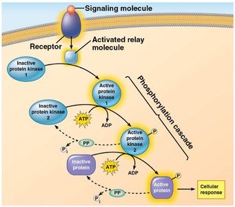
Second Messengers
Small, nonprotein molecules or ions that spread signals inside the cell. - Common second messengers: cAMP (cyclic AMP), Ca2+ ions. - Pathways: Both GPCRs and RTKs can use second messengers. - cAMP: Produced from ATP by adenylyl cyclase; degraded to AMP by phosphodiesterase. 
cAMP in G Protein Pathways
- Step 1: First messenger binds GPCR. - Step 2: GPCR activates G protein. - Step 3: G protein activates adenylyl cyclase. - Step 4: Adenylyl cyclase converts ATP to cAMP. - Step 5: cAMP activates protein kinase A, triggering cellular responses. 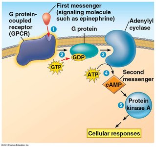
Cellular Responses
Nuclear Responses
Many signaling pathways regulate gene expression. - Transcription factors: Activated by signaling cascades, turn genes on or off. - Example: Growth factors activate specific genes for cell growth. 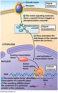
Cytoplasmic Responses
Some pathways regulate enzyme activity or metabolic processes. - Example: Epinephrine stimulates glycogen breakdown in muscle cells, providing glucose for energy. 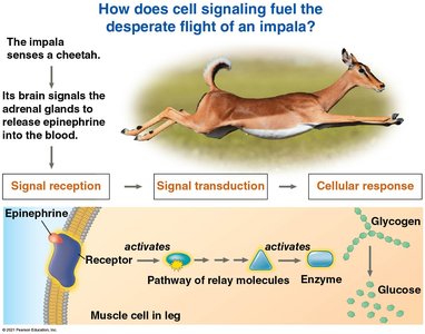
Signal Amplification and Specificity
Amplification
A single signaling molecule can produce a large cellular response. - Mechanism: Each step in the pathway activates multiple molecules of the next component. - Example: One epinephrine molecule can lead to the production of millions of glucose molecules. 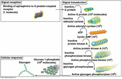
Specificity
Only cells with the appropriate receptor respond to a given messenger. - Different responses: The same signal can trigger different outcomes in different cell types, depending on their internal proteins and receptors.
Resetting the Signal: Termination
Termination Mechanisms
To respond to new signals, activated components must be switched off. - Ligand leaves receptor: Binding is reversible. - G protein turns off: GTP hydrolyzed to GDP. - cAMP removed: Phosphodiesterase converts cAMP to AMP. - Proteins dephosphorylated: Protein phosphatases remove phosphate groups. - Ion channels close: Ions are pumped out of the cytosol.
Case Study: Insulin and Diabetes
Insulin Signaling and Glucose Regulation
Insulin is a peptide hormone that enables glucose uptake into cells. - Normal function: Insulin binds its receptor, triggering pathways that allow glucose to enter muscle and liver cells, where it is stored as glycogen. - Diabetes: Occurs when the body does not produce enough insulin or cannot use it properly, leading to high blood sugar levels. 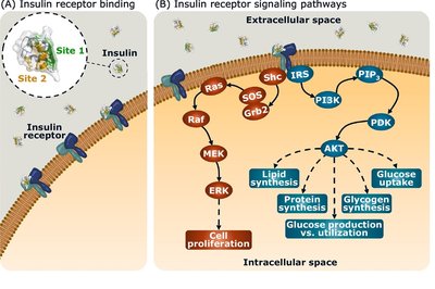
Insulin Receptor Signaling Pathways
Insulin binding activates multiple intracellular pathways, affecting lipid synthesis, protein synthesis, glycogen synthesis, and glucose uptake.
Summary Table: Cell Signaling Pathways
Step | Key Event | Example Molecule |
|---|---|---|
Reception | Ligand binds receptor | Epinephrine, insulin |
Transduction | Relay molecules, second messengers, kinase cascades | G protein, cAMP, protein kinase A |
Response | Gene expression, enzyme activation, metabolic change | Glycogen breakdown, glucose uptake |
Reset | Signal termination, return to resting state | Phosphodiesterase, protein phosphatases |
Additional info: These notes expand on the original content by providing definitions, examples, and context for each step and mechanism in cell signaling, making the material self-contained and suitable for exam preparation.
