 Indietro
Cell Structure and Function: A Mini-Textbook Study Guide
Guida di studio - Note intelligenti
 Appunti personalizzati basati sui tuoi materiali, ampliati con definizioni chiave, esempi e contesto.
Cell Theory
Foundations of Cell Theory
The cell theory, proposed by Schleiden and Schwann in 1839, is a fundamental concept in biology that describes the properties of cells as the basic unit of life.
All organisms are composed of cells: Every living thing is made up of one or more cells.
Cells are the smallest living things: The cell is the basic structural and functional unit of life.
Cells arise only from pre-existing cells: New cells are produced by the division of existing cells.
Continuous line of descent: All cells today trace their lineage back to the first living cells.
Surface Area-to-Volume Ratio
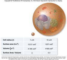
As a cell increases in size, its volume grows much faster than its surface area. This ratio is critical for cell function, as it affects the ability of the cell to exchange materials with its environment.
Surface area-to-volume ratio: Volume increases 10 times faster than surface area as cell size increases.
Implications: Larger cells have less surface area relative to their volume, limiting efficient transport of materials.
Formula:
Cell Size and Microscopy
Scale of Biological Structures
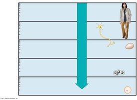
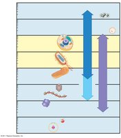
Cells and their components vary greatly in size, from atoms and molecules to entire cells visible to the naked eye. Microscopy techniques allow us to visualize these structures at different scales.
Microscopy: Light, electron, and super-resolution microscopy are used to observe cells and their organelles.
Cell size: Most plant and animal cells are 10–100 μm in diameter; some cells (e.g., eggs, nerve cells) are much larger.
Basic Cell Structures
Common Features of All Cells
Despite their diversity, all cells share certain structural features essential for life.
Genetic material: Located in a nucleoid (prokaryotes) or nucleus (eukaryotes).
Cytoplasm: Semifluid matrix filling the cell.
Plasma membrane: Phospholipid bilayer that encloses the cell.
Ribosomes: Structures for protein synthesis.
Prokaryotic vs. Eukaryotic Cells
Prokaryotic Cells
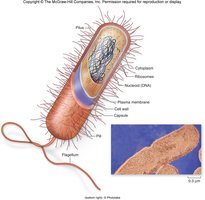
Prokaryotic cells are simpler and lack membrane-bound organelles. They are found in the domains Bacteria and Archaea.
No nucleus: DNA is located in an unbound region called the nucleoid.
No membrane-bound organelles: All cellular processes occur in the cytoplasm.
Cell wall: Provides structural support.
Ribosomes: Present for protein synthesis.
Eukaryotic Cells
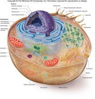
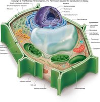
Eukaryotic cells are more complex, with internal membranes that compartmentalize functions. They are found in protists, fungi, animals, and plants.
Nucleus: Contains most of the cell's DNA.
Membrane-bound organelles: Specialized structures for various functions.
Compartmentalization: Allows for efficient and specialized cellular processes.
Plasma Membrane Structure and Function
Phospholipid Bilayer
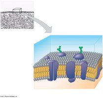
The plasma membrane is a selective barrier composed of a phospholipid bilayer with embedded proteins and carbohydrates. It regulates the passage of substances in and out of the cell.
Hydrophilic regions: Face outward toward the aqueous environment.
Hydrophobic regions: Face inward, away from water.
Proteins: Serve as channels, receptors, and enzymes.
Carbohydrate side chains: Involved in cell recognition.
Nucleus and Ribosomes
Nucleus
The nucleus is the control center of the cell, housing most of the genetic material and coordinating activities such as growth and reproduction.
Nucleolus: Manufactures ribosomal subunits.
Nuclear envelope: Double membrane with pores for material exchange.
Ribosomes
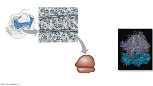
Ribosomes are the sites of protein synthesis, composed of ribosomal RNA and protein. They can be free in the cytosol or bound to the endoplasmic reticulum.
Free ribosomes: Synthesize proteins for use within the cell.
Bound ribosomes: Synthesize proteins for secretion or membrane insertion.
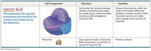
Cell Component | Structure | Function |
|---|---|---|
Nucleus | Surrounded by nuclear envelope; contains nucleolus | Houses chromosomes (DNA); nucleolus makes ribosomal subunits |
Ribosome | Two subunits made of rRNA and protein; free or bound | Protein synthesis |
Endomembrane System
Components and Functions
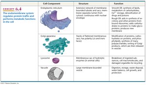
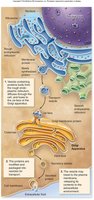
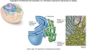
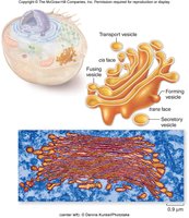
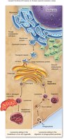
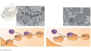
The endomembrane system is a series of membranes that divide the cell into compartments, facilitating specialized functions.
Endoplasmic reticulum (ER): Rough ER synthesizes proteins; smooth ER synthesizes lipids and detoxifies chemicals.
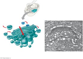
Golgi apparatus: Modifies, sorts, and packages proteins and lipids for transport.
Lysosomes: Digest macromolecules and recycle cellular components.
Vacuoles: Storage and maintenance compartments, especially prominent in plant cells.
Cell Component | Structure | Function |
|---|---|---|
Endoplasmic reticulum | Network of membranous tubules and sacs | Protein and lipid synthesis, detoxification |
Golgi apparatus | Stacks of flattened membranes | Modification, sorting, packaging of proteins and lipids |
Lysosome | Membranous sac of hydrolytic enzymes | Digestion of macromolecules |
Vacuole | Large membrane-bounded vesicle | Storage, waste disposal, water regulation |
Endosymbiotic Organelles
Mitochondria and Plastids
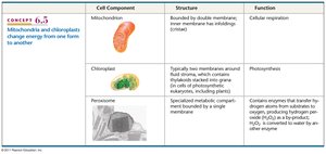
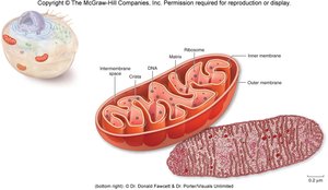
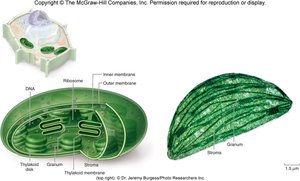
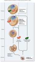
Mitochondria and plastids (chloroplasts) are organelles derived from free-living bacteria that became integrated into eukaryotic cells through endosymbiosis.
Mitochondria: Site of cellular respiration; converts energy from food into ATP.
Chloroplasts: Site of photosynthesis in plant cells.
Cell Component | Structure | Function |
|---|---|---|
Mitochondrion | Double membrane; inner membrane has infoldings (cristae) | Cellular respiration |
Chloroplast | Double membrane; contains thylakoids and stroma | Photosynthesis |
Peroxisome | Specialized metabolic compartment | Breakdown of hydrogen peroxide and other metabolic functions |
Cytoskeleton
Structure and Function
The cytoskeleton is a network of protein fibers that provides structural support, maintains cell shape, and facilitates movement of organelles and materials within the cell.
Actin filaments: Responsible for cellular contractions, crawling, and pinching.
Microtubules: Provide organization and move materials within the cell.
Intermediate filaments: Provide structural stability.
Cell Movement
Mechanisms of Cell Movement
Cells move using various mechanisms, including actin-myosin interactions, flagella, and cilia.
Crawling: Achieved via actin filaments and myosin.
Flagella: Undulate to propel cells.
Cilia: Arranged in rows to move cells or substances across cell surfaces.
Extracellular Structures
Extracellular Matrix (ECM)
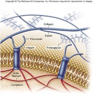
The extracellular matrix surrounds animal cells and is composed of glycoproteins and fibrous proteins such as collagen. It provides structural support and can be connected to the cytoplasm via integrin proteins.
Collagen: Provides tensile strength.
Elastin: Provides elasticity.
Fibronectin and proteoglycans: Aid in cell adhesion and signaling.
Integrins: Connect ECM to the cytoskeleton.
Summary Table: Cell Components and Functions
Cell Component | Structure | Function |
|---|---|---|
Nucleus | Double membrane, nucleolus | Genetic information storage, ribosome assembly |
Ribosome | rRNA and protein, free or bound | Protein synthesis |
ER | Membranous tubules, rough and smooth | Protein and lipid synthesis |
Golgi apparatus | Flattened sacs | Modification, sorting, packaging |
Lysosome | Hydrolytic enzymes | Digestion, recycling |
Vacuole | Large vesicle | Storage, waste disposal |
Mitochondrion | Double membrane, cristae | Cellular respiration |
Chloroplast | Double membrane, thylakoids | Photosynthesis |
Peroxisome | Single membrane | Metabolic breakdown |
Conclusion
Understanding cell structure and function is fundamental to biology. Cells are the basic unit of life, and their organization, compartmentalization, and specialized structures enable the diverse functions necessary for living organisms.
