 Indietro
Cell Structure and Function: A Tour of the Cell
Guida di studio - Note intelligenti
 Appunti personalizzati basati sui tuoi materiali, ampliati con definizioni chiave, esempi e contesto.
Overview: The Fundamental Units of Life
Introduction to Cells
Cells are the basic structural and functional units of all living organisms. Every organism is composed of one or more cells, and cell structure is closely linked to cellular function. All cells share a common ancestry, tracing back to earlier cells.
Cell: The simplest collection of matter that can live.
Cell structure-function relationship: The shape and internal organization of a cell are adapted to its specific functions.
Cell lineage: All cells arise from pre-existing cells.
Microscopy and Cell Study
Microscopes in Biology
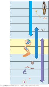
Biologists use various types of microscopes to study cells and their components.
Light Microscope (LM): Uses visible light and glass lenses to magnify specimens. Suitable for viewing most plant and animal cells, bacteria, and some organelles.
Electron Microscopes (EM): Use electron beams for much higher resolution.
Scanning Electron Microscope (SEM): Provides detailed 3-D images of cell surfaces.
Transmission Electron Microscope (TEM): Reveals internal cell structures.
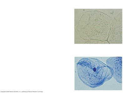
Microscope Techniques
Brightfield (unstained): Shows cells in their natural state.
Brightfield (stained): Enhances contrast to reveal cell structures.
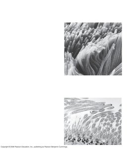
Electron Microscopy
SEM: Used for surface details.
TEM: Used for internal details.
Comparing Prokaryotic and Eukaryotic Cells
Basic Features of All Cells
All cells, whether prokaryotic or eukaryotic, share certain features:
Plasma membrane: Selective barrier for substances entering or leaving the cell.
Cytoplasm: Semifluid substance inside the cell.
DNA: Genetic material.
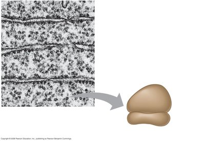
Ribosomes: Sites of protein synthesis.
Prokaryotic vs. Eukaryotic Cells
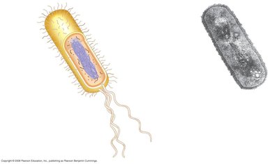
Prokaryotic cells:
No nucleus; DNA is in the nucleoid region.
No membrane-bound organelles.
Domains: Bacteria and Archaea.
Eukaryotic cells:
DNA enclosed in a nucleus.
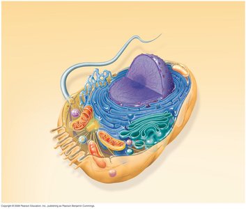
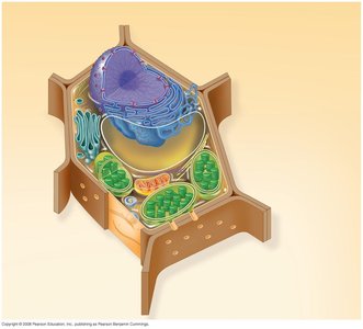
Membrane-bound organelles (e.g., mitochondria, ER).
Domains: Protists, fungi, animals, plants.
Generally larger than prokaryotic cells.
Cellular Structures and Organelles
Plasma Membrane
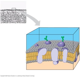
The plasma membrane is a phospholipid bilayer with embedded proteins and carbohydrates, acting as a selective barrier.
Structure: Double layer of phospholipids.
Function: Regulates passage of oxygen, nutrients, and waste.
Nucleus and Ribosomes
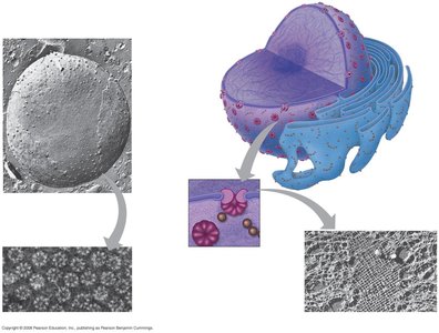
Nucleus: Contains most of the cell's DNA; surrounded by a nuclear envelope with pores.
Nucleolus: Site of ribosome synthesis.
Ribosomes: Made of rRNA and protein; carry out protein synthesis.
Free ribosomes: In cytosol; synthesize proteins for use within the cell.
Bound ribosomes: Attached to ER or nuclear envelope; synthesize proteins for export or membrane insertion.
Endomembrane System
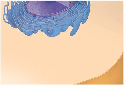
The endomembrane system regulates protein traffic and performs metabolic functions. Components include:
Nuclear envelope
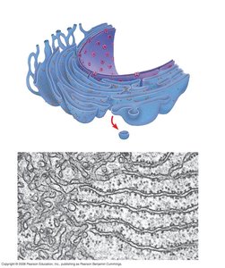
Endoplasmic reticulum (ER):
Smooth ER: Synthesizes lipids, metabolizes carbohydrates, detoxifies poisons, stores calcium.
Rough ER: Has bound ribosomes; synthesizes glycoproteins, distributes transport vesicles, produces membranes.
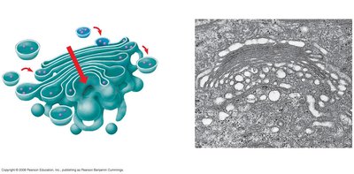
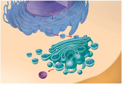
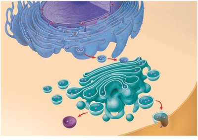
Golgi apparatus: Modifies, sorts, and packages proteins and lipids.
Lysosomes: Digestive compartments with hydrolytic enzymes.
Vacuoles: Storage and maintenance compartments.
Plasma membrane
Lysosomes and Vacuoles
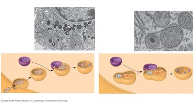
Lysosomes: Digest macromolecules; involved in phagocytosis and autophagy.
Vacuoles:
Food vacuoles: Formed by phagocytosis.
Contractile vacuoles: Pump excess water out.
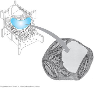
Central vacuole: Stores water and organic compounds in plants.
Mitochondria and Chloroplasts
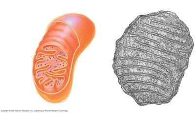
Mitochondria: Sites of cellular respiration; generate ATP; have double membrane and their own DNA.
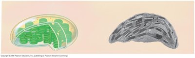
Chloroplasts: Sites of photosynthesis; contain chlorophyll; found in plants and algae; have double membrane and their own DNA.
The Cytoskeleton
Structure and Function
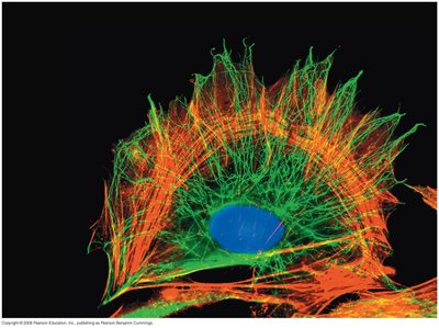
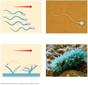
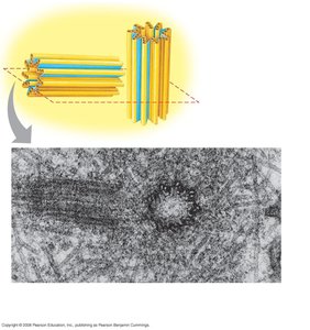
The cytoskeleton is a network of fibers that organizes cell structures and activities, providing support, motility, and regulation.
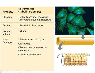
Microtubules: Thickest; made of tubulin; involved in cell shape, motility, chromosome movement, and organelle transport.
Microfilaments (Actin filaments): Thinnest; made of actin; involved in cell shape, muscle contraction, cytoplasmic streaming, and cell motility.
Intermediate filaments: Middle diameter; made of keratin; provide structural support.
Extracellular Components and Cell Junctions
Cell Walls and Extracellular Matrix (ECM)
Cell wall: Found in plants, fungi, prokaryotes, and some protists; provides protection, shape, and prevents water uptake; made of cellulose and other polysaccharides.
ECM: Found in animal cells; composed of glycoproteins (e.g., collagen, proteoglycans); provides structural support and cell signaling.
Intercellular Junctions
Plasmodesmata: Channels between plant cells for transport of water, solutes, proteins, and RNA.
Tight junctions: Seal neighboring animal cells to prevent leakage.
Desmosomes: Anchor animal cells together into sheets.
Gap junctions: Provide cytoplasmic channels for communication between animal cells.
Summary Table: Differences Between Prokaryotic and Eukaryotic Cells
Feature | Prokaryotic Cells | Eukaryotic Cells |
|---|---|---|
Nucleus | No (nucleoid region) | Yes (membrane-bound) |
Organelles | No membrane-bound organelles | Membrane-bound organelles present |
Size | Smaller (0.1–5 µm) | Larger (10–100 µm) |
Domains | Bacteria, Archaea | Protists, Fungi, Animals, Plants |
Summary Table: Differences Between Plant and Animal Cells
Feature | Plant Cells | Animal Cells |
|---|---|---|
Cell Wall | Present (cellulose) | Absent |
Chloroplasts | Present | Absent |
Central Vacuole | Large, present | Small or absent |
Lysosomes | Rare | Common |
Centrioles | Absent | Present |
Key Functions of Eukaryotic Organelles
Nucleus: Stores genetic information.
Ribosomes: Protein synthesis.
ER: Synthesis of lipids (smooth), proteins (rough).
Golgi apparatus: Modification, sorting, packaging of proteins and lipids.
Lysosomes: Digestion of macromolecules.
Vacuoles: Storage and maintenance.
Mitochondria: ATP production.
Chloroplasts: Photosynthesis.
Cytoskeleton: Support, motility, regulation.
Cell junctions: Communication and adhesion.
ECM: Structural support and signaling.
Additional info:
Cell size is limited by surface-to-volume ratio, which affects the efficiency of material exchange.
Endosymbiont theory explains the origin of mitochondria and chloroplasts as formerly free-living prokaryotes engulfed by ancestral eukaryotic cells.
