 Indietro
Cell Structure and Function: A Tour of the Cell
Guida di studio - Note intelligenti
 Appunti personalizzati basati sui tuoi materiali, ampliati con definizioni chiave, esempi e contesto.
Cell Structure and Function
Overview of Cell Types
Cells are the fundamental units of life, classified into two main types: prokaryotic and eukaryotic. Prokaryotes include Bacteria and Archaea, while eukaryotes encompass Protists, Fungi, Plants, and Animals. The distinction between these cell types is based on the presence or absence of a nucleus and membrane-bound organelles.
Prokaryotic cells: Lack a nucleus; DNA is located in the nucleoid region.
Eukaryotic cells: Possess a nucleus and various organelles.
Domains: Bacteria, Archaea (prokaryotes); Eukarya (eukaryotes).
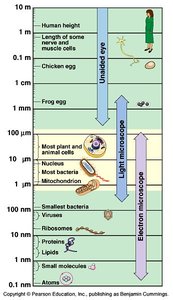
Cell Size and Microscopy
Cell size varies widely, from microscopic bacteria to large eukaryotic cells. The ability to observe cells depends on the type of microscope used. Light microscopes are suitable for most plant and animal cells, while electron microscopes are required for smaller structures such as ribosomes and viruses.
Microscopy: Light microscopes for cells; electron microscopes for organelles and molecules.
Scale: Cells range from 1–100 µm; organelles and molecules are smaller.
Prokaryotic Cells
Structure of Prokaryotic Cells
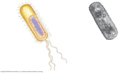
Prokaryotic cells are characterized by their simplicity and lack of membrane-bound organelles. They possess a plasma membrane, cell wall, capsule, and sometimes structures such as fimbriae and flagella for movement and attachment.
Nucleoid: Region containing DNA.
Ribosomes: Sites of protein synthesis.
Cell wall: Provides structural support.
Capsule: Offers protection and aids in adhesion.
Flagella: Enables motility.
Archaea
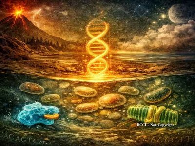
Archaea are single-celled prokaryotes with unique genome organization, ribosome structure, and metabolic pathways that are more closely related to eukaryotes than to bacteria. They are grouped in their own domain due to these distinct features.
Unique features: Genome and ribosome similarities to eukaryotes.
Domain: Archaea.
Eukaryotic Cells
Structure of Eukaryotic Cells
Eukaryotic cells are complex, containing a nucleus and various organelles. Animal and plant cells share many features but also have unique structures.
Nucleus: Contains DNA, surrounded by a nuclear envelope.
Organelles: Endoplasmic reticulum (ER), Golgi apparatus, mitochondria, lysosomes, peroxisomes.
Animal cells: Have centrioles, lysosomes, flagella.
Plant cells: Have chloroplasts, central vacuole, cell wall, plasmodesmata.
Cell Walls and Membranes
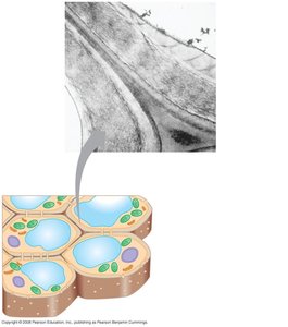
Plant cells are surrounded by a cell wall composed of cellulose, providing structural support. Both plant and animal cells have a plasma membrane, a phospholipid bilayer with embedded proteins and carbohydrates.
Cell wall: Primary and secondary walls, middle lamella, plasmodesmata.
Plasma membrane: Hydrophilic and hydrophobic regions, proteins, carbohydrates.
The Cytoskeleton
Components of the Cytoskeleton
The cytoskeleton is a network of protein filaments that provides structural support, enables cell movement, and organizes organelles. It consists of microtubules, microfilaments, and intermediate filaments.
Microtubules: Hollow tubes, originate from centrosomes, mediate movement of organelles and chromosomes.
Microfilaments (Actin): Thin filaments, determine cell shape, involved in cell division and muscle contraction.
Intermediate filaments: Provide structural integrity, anchor cell components.
Microtubules
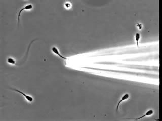
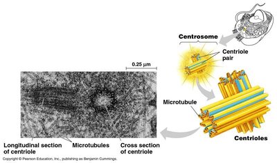
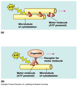
Microtubules are dynamic structures that originate from centrosomes and are involved in cellular transport, chromosome movement, and cell motility. Motor proteins such as dyneins and kinesins use ATP to move along microtubules.
Centrosome: Microtubule organizing center.
Motor proteins: Dyneins and kinesins.
Functions: Organelle movement, chromosome separation, cell motility (flagella and cilia).
Microfilaments (Actin)
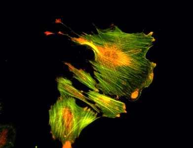
Microfilaments are composed of actin and are essential for maintaining cell shape, enabling membrane movement, and facilitating muscle contraction and cytoplasmic streaming.
Actin: Protein forming microfilaments.
Functions: Cell shape, division, muscle contraction, cytoplasmic streaming.
Intermediate Filaments
Intermediate filaments provide structural integrity and anchor cell components. Examples include lamins and keratins.
Lamins: Nuclear envelope support.
Keratins: Structural proteins in epithelial cells.
Cellular Organelles
Ribosomes
Ribosomes are responsible for protein synthesis and can be found free in the cytoplasm or bound to the endoplasmic reticulum.
Free ribosomes: Synthesize proteins for use within the cell.
Bound ribosomes: Synthesize proteins for secretion or membrane insertion.
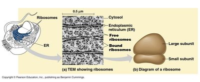
Endoplasmic Reticulum (ER)
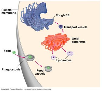
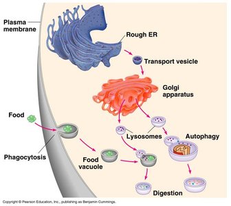
The ER is a network of membranes involved in protein and lipid synthesis. The rough ER is studded with ribosomes, while the smooth ER lacks ribosomes and is involved in lipid metabolism.
Rough ER: Protein synthesis and processing.
Smooth ER: Lipid synthesis and detoxification.
Golgi Apparatus
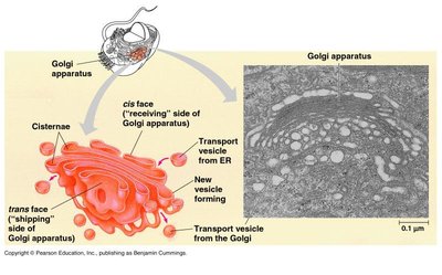
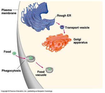
The Golgi apparatus modifies, sorts, and packages proteins and lipids for secretion or delivery to other organelles.
Cisternae: Flattened membrane sacs.
cis face: Receiving side.
trans face: Shipping side.
Lysosomes
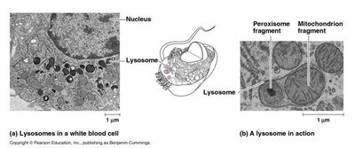
Lysosomes are the major site for digestion of external compounds and cellular debris. They contain hydrolytic enzymes that break down macromolecules.
Function: Digestion of food, autophagy, recycling of cellular components.
Enzymes: Hydrolytic enzymes.
Mitochondria
Mitochondria are the site of ATP synthesis in both plant and animal cells. They are often referred to as the "powerhouse" of the cell.
Function: Cellular respiration and energy production.
Chloroplasts
Chloroplasts are found only in plant cells and are responsible for photosynthesis, converting light energy into chemical energy.
Function: Photosynthesis.
Extracellular Structures and Cell Junctions
Extracellular Matrix (ECM)
The ECM is a network of proteins and carbohydrates outside the cell that provides structural support and mediates cell signaling.
Components: Collagen, proteoglycans, fibronectin.
Function: Support, adhesion, communication.
Cell Junctions
Cells are held together by specialized junctions that facilitate communication and maintain tissue integrity.
Tight junctions: Prevent fluid movement between cells.
Desmosomes: Provide mechanical strength.
Gap junctions: Allow passage of ions and small molecules.
Summary Table: Comparison of Cell Types
Feature | Prokaryotic Cell | Eukaryotic Cell |
|---|---|---|
Nucleus | No | Yes |
Organelles | No | Yes |
Cell Wall | Yes (most) | Yes (plants, fungi) |
DNA Location | Nucleoid | Nucleus |
Size | 1–10 µm | 10–100 µm |
Examples | Bacteria, Archaea | Plants, Animals, Fungi, Protists |
Key Equations
Surface area to volume ratio is critical for cell function:
*Additional info: Expanded explanations and context were added to ensure completeness and clarity for exam preparation.*
