 Indietro
Cell Structure and Function: Foundations of Cellular Biology
Guida di studio - Note intelligenti
 Appunti personalizzati basati sui tuoi materiali, ampliati con definizioni chiave, esempi e contesto.
Cell Structure and Function
Introduction to Cells and Biomolecules
Cells are the fundamental units of life, composed of four major classes of biomolecules: nucleic acids, proteins, carbohydrates, and lipids. These molecules form the structural and functional basis of all living organisms.
Nucleic acids: Store and transmit genetic information (DNA and RNA).
Proteins: Perform a wide range of functions including catalysis, structure, and transport.
Carbohydrates: Provide energy and structural support.
Lipids: Form membranes and store energy.
Cells can be observed under a microscope, revealing their complex internal structures.
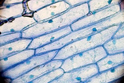
History of Cell Discovery
The development of the microscope in the late 16th and 17th centuries led to the discovery of cells and the formulation of cell theory. Key contributors include:
Robert Hooke (1665): First to describe cells in cork tissue.
Anton van Leeuwenhoek (1674): Observed unicellular organisms.
Matthias Schleiden (1838): Proposed that plants are made of cells.
Theodor Schwann (1839): Proposed that animals are made of cells.

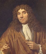
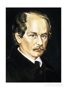
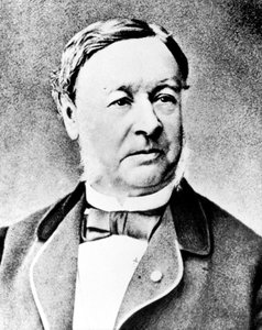
Cell Theory
Cell theory is a foundational concept in biology, stating:
All living organisms are composed of one or more cells.
The cell is the basic structural and functional unit of life.
All cells arise from pre-existing cells by division.
Cells contain hereditary material passed to offspring during division.
Types of Cells: Prokaryotic and Eukaryotic
Prokaryotic Cells
Prokaryotic cells are the simplest forms of life, lacking a nucleus and membrane-bound organelles. They include Bacteria and Archaea.
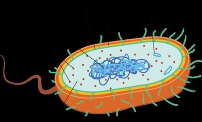
Components: plasma membrane, cytosol/cytoplasm, genetic material (chromosomes), and ribosomes.
Prokaryotes are characterized by their simplicity and efficiency.
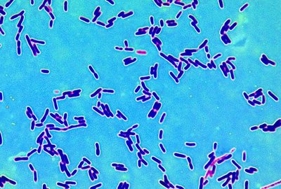
Prokaryotic Diversity
Prokaryotes are divided into two domains: Bacteria and Archaea. Archaea often inhabit extreme environments, such as hot springs.
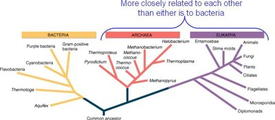
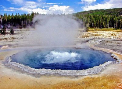
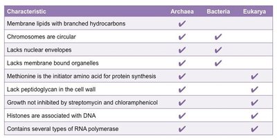
Comparison of Prokaryotic Domains
Characteristic | Archaea | Bacteria | Eukarya |
|---|---|---|---|
Membrane lipids with branched hydrocarbons | ✓ | ||
Chromosomes are circular | ✓ | ✓ | |
Lacks nuclear envelopes | ✓ | ✓ | |
Lacks membrane-bound organelles | ✓ | ✓ | |
Methionine initiator for protein synthesis | ✓ | ✓ | |
Lack peptidoglycan in cell wall | ✓ | ✓ | |
Growth not inhibited by streptomycin/chloramphenicol | ✓ | ✓ | |
Histones associated with DNA | ✓ | ✓ | |
Several types of RNA polymerase | ✓ | ✓ |
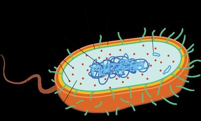
Prokaryotic Cell Structure
Capsule: Sticky layer for adhesion and protection.
Cell Wall: Provides shape and protection; permeable to water.
Plasma Membrane: Phospholipid bilayer controlling entry/exit of substances.
Pili: Surface appendages for attachment or DNA exchange.
Flagella: Used for motility.
Ribosomes: Sites of protein synthesis.
Plasmids: Small, circular DNA molecules with extra genes.
Nucleoid: Region containing the main circular DNA molecule.
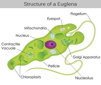
Eukaryotic Cells
Eukaryotic cells are more complex, containing internal membranes and specialized organelles. They can be autotrophic (plant-like) or heterotrophic (animal-like).
Internal membranes compartmentalize functions and increase surface area for reactions.
Examples: plants, animals, fungi, and protists.
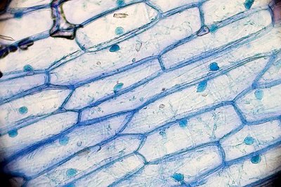
Plant-like (Autotrophic) and Animal-like (Heterotrophic) Cells
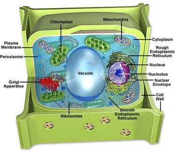
Plant-like cells: Contain chloroplasts, large central vacuole, and cell wall.
Animal-like cells: Contain centrioles, lysosomes, and lack a cell wall.
Cell Differentiation
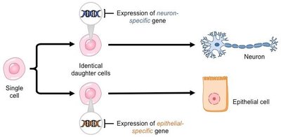
In multicellular organisms, cells differentiate to perform specialized functions, resulting in diverse tissues and organs.
Endomembrane System and Organelles
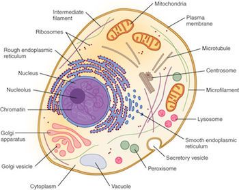
Nucleus and Nucleolus
The nucleus stores genetic material and is the site of DNA replication and transcription. The nucleolus, a region within the nucleus, produces ribosomes.
Nucleus: Contains DNA, controls cell activities.
Nucleolus: Synthesizes ribosomal RNA and assembles ribosomes.
Ribosomes
Ribosomes are the sites of protein synthesis. Free ribosomes produce cytoplasmic proteins, while bound ribosomes (on rough ER) synthesize membrane and secretory proteins.
Endoplasmic Reticulum (ER)
Rough ER: Studded with ribosomes; synthesizes and processes proteins.
Smooth ER: Lacks ribosomes; synthesizes lipids, detoxifies chemicals, and stores calcium.
Golgi Apparatus
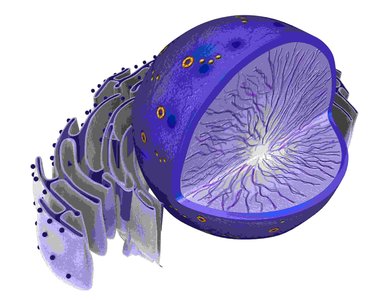
The Golgi apparatus modifies, sorts, and packages proteins and lipids for secretion or delivery to other organelles.
Vesicles and Vacuoles
Vesicles: Small membrane-bound sacs for transport and storage.
Vacuoles: Large vesicles for storage; central vacuole in plants maintains turgor pressure.
Contractile vacuoles: Expel excess water in some protists.
Lysosomes and Peroxisomes
Lysosomes: Contain hydrolytic enzymes for digestion of macromolecules and old organelles; found mainly in animal cells.
Peroxisomes: Break down fatty acids and detoxify harmful substances; found in nearly all eukaryotes.
Energy Organelles: Mitochondria and Chloroplasts
Mitochondria: Site of aerobic respiration and ATP production; contain their own DNA and ribosomes.
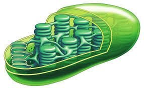
Chloroplasts: Site of photosynthesis in plants and algae; contain chlorophyll, thylakoids, grana, and their own DNA.
Endosymbiotic Theory
Mitochondria and chloroplasts are believed to have originated from free-living prokaryotes that entered into a symbiotic relationship with ancestral eukaryotic cells. Evidence includes their own DNA and double membranes.
Cytoskeleton
The cytoskeleton is a network of protein filaments that provides structural support, enables cell movement, and organizes organelles.
Microtubules: Hollow tubes for cell shape, transport, and division.
Microfilaments: Thin filaments for movement and shape changes.
Intermediate filaments: Provide mechanical support.
External Cellular Structures
Cell Wall: Provides support and protection; made of cellulose in plants, chitin in fungi, and peptidoglycan in bacteria.
Extracellular Matrix (ECM): Network of proteins (e.g., collagen) outside animal cells for support and adhesion.
Cell Junctions: Protein structures that connect cells and facilitate communication.
Cell Membrane Structure and Function
Phospholipid Bilayer and Fluid Mosaic Model
The cell membrane is composed of a double layer of phospholipids with embedded proteins, cholesterol, and carbohydrates. This structure is described by the fluid mosaic model.
Phospholipids: Amphipathic molecules with hydrophilic heads and hydrophobic tails.
Proteins: Peripheral (surface) and integral (embedded) proteins serve roles in transport, signaling, and structure.
Cholesterol: Maintains membrane fluidity in animal cells.
Carbohydrates: Glycoproteins and glycolipids for cell recognition and communication.
Membrane Transport
Passive Transport: Movement of substances down their concentration gradient without energy input.
Simple diffusion: Small, uncharged molecules (e.g., O2, CO2).
Facilitated diffusion: Large or charged molecules via membrane proteins.
Osmosis: Diffusion of water across a semipermeable membrane.
Active Transport: Movement of substances against their concentration gradient, requiring ATP (e.g., Na+/K+ pump).
Bulk Transport: Endocytosis (phagocytosis, pinocytosis) and exocytosis for large particles or volumes.
Osmosis and Tonicity
Isotonic: Equal solute concentration; no net water movement.
Hypotonic: Lower solute concentration outside; water enters cell.
Hypertonic: Higher solute concentration outside; water leaves cell.
In plant cells, turgor pressure from the central vacuole maintains structure. In animal cells, extreme tonicity can cause lysis or crenation.
Water Potential
Water potential (ψ) determines the direction of water movement. Water moves from areas of higher (less negative) to lower (more negative) water potential, influenced by solute concentration and pressure.
Equation:
= total water potential
= solute potential
= pressure potential
