 Indietro
Cell Structure and Function: Study Notes for General Biology
Guida di studio - Note intelligenti
 Appunti personalizzati basati sui tuoi materiali, ampliati con definizioni chiave, esempi e contesto.
Cell Structure and Function
Historical Foundations of Cell Biology
The discovery of cells and the development of cell theory are foundational to modern biology. Early observations using microscopes led to the identification of cells as the basic unit of life.
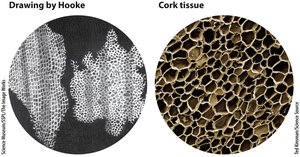
Robert Hooke (1665): Used a simple light microscope to observe cork, identifying 'cells' as compartments resembling monks' rooms. He observed dead cells, seeing only cell walls and empty spaces.
Microscope: The invention and improvement of microscopes enabled scientists to observe cells and their structures.
Cork Tissue: Hooke's drawings of cork tissue revealed the cellular structure, confirming the existence of cells.



7.1: The Cell Theory
Cell theory is a fundamental concept in biology, describing the properties and significance of cells.
All organisms are made up of cells.
Cells are the basic unit of living things.
All cells come from pre-existing cells.
Organelle: Specialized structures within a cell, each with specific functions (e.g., mitochondria, nucleus).
7.2: Cell Membrane
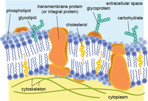
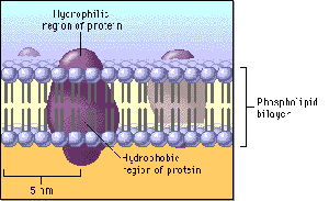
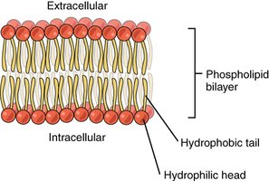
The cell membrane, or plasma membrane, is a dynamic structure that separates the cell from its environment and regulates the movement of substances.
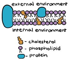
Structure: Composed of a phospholipid bilayer with embedded proteins and cholesterol.
Function: Maintains cellular integrity, controls entry and exit of materials.
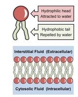
Phospholipid Bilayer: Hydrophilic heads face outward; hydrophobic tails face inward.
Cholesterol: Maintains membrane fluidity and flexibility.
7.3: All Cells
All cells share certain essential components that are critical for their function and survival.
Cytoplasm: Aqueous solution inside the plasma membrane, containing organelles and molecules.
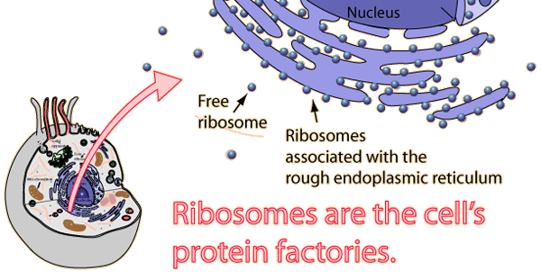
Ribosomes: Small, circular structures responsible for protein synthesis.
Universal Features: All cells possess a plasma membrane, cytoplasm, and ribosomes.
7.4: Eukaryotic vs. Prokaryotic Cells
Cells are classified as either eukaryotic or prokaryotic based on their structural complexity and presence of organelles.
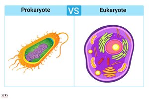
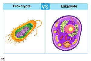
Eukaryotic Cells: Complex, contain a nucleus, larger size, DNA in long strands. Examples: plant and animal cells.
Prokaryotic Cells: Simple, lack a nucleus, smaller size, circular DNA. Example: bacteria.
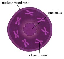
Nucleus: Membrane-bound organelle housing genetic material (DNA), controls cell activities.
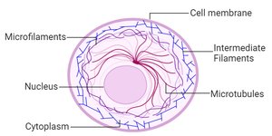
Cytoskeleton: Network of protein filaments providing structural support and shape.
7.5: Plant vs. Animal Cells
Plant and animal cells share many features but also have distinct differences in their organelles and structures.
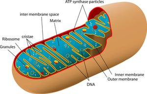
Mitochondria: Double-membraned organelle, site of cellular respiration and energy production. Known as the 'powerhouse' of the cell.
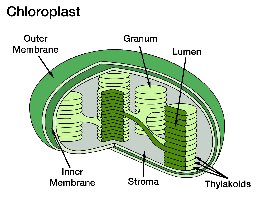
Chloroplasts: Double-membraned organelle in plant cells, site of photosynthesis, contains green pigments (chlorophyll).
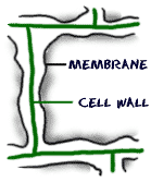
Cell Wall: Rigid structure made of cellulose, provides support and protection in plant cells.
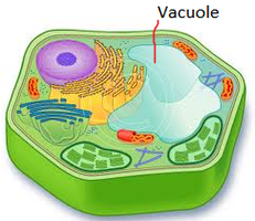
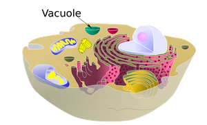
Vacuole: Membrane-bound sac for storage of nutrients and waste; large in plant cells, smaller and more numerous in animal cells.
Key Differences: Plant cells have chloroplasts, cell walls, and a large central vacuole; animal cells lack chloroplasts and cell walls, and have multiple small vacuoles.
7.6: Membrane Organelles
Membrane-bound organelles are specialized structures within eukaryotic cells, each with unique functions.
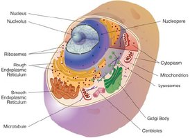
Endoplasmic Reticulum (ER): Network of membranes near the nucleus; rough ER has ribosomes and synthesizes proteins, smooth ER lacks ribosomes and synthesizes lipids.
Vesicle: Small membrane sphere for transport of materials within the cell.
Golgi Body (Apparatus): Stack of membrane discs that modify, store, and route proteins and other molecules.
Lysosome: Membrane-bound container with digestive enzymes, breaks down food, damaged organelles, and dead cells.
Summary Table: Plant vs. Animal Cell Features
Feature | Plant Cell | Animal Cell |
|---|---|---|
Chloroplasts | Present | Absent |
Mitochondria | Present | Present |
Cell Wall | Present | Absent |
Vacuole | One large | Many small |
Key Terms and Definitions
Cell: Basic unit of life, enclosed by a membrane.
Organelle: Specialized structure within a cell.
Plasma Membrane: Lipid bilayer separating cell from environment.
Cytoplasm: Fluid inside cell containing organelles.
Ribosome: Protein-synthesizing structure.
Nucleus: Organelle containing DNA.
Cytoskeleton: Network of proteins for support.
Mitochondria: Organelle for energy production.
Chloroplast: Organelle for photosynthesis.
Cell Wall: Rigid structure in plant cells.
Vacuole: Storage organelle.
Endoplasmic Reticulum: Membrane network for synthesis.
Golgi Body: Organelle for modification and transport.
Lysosome: Organelle for digestion.
Key Formula: Surface Area to Volume Ratio
The surface area to volume ratio is important for cell function, affecting transport and communication.
Formula: For a sphere, surface area = , volume =
Ratio:
Example: Smaller cells have a higher surface area to volume ratio, facilitating efficient transport of materials.
