 Indietro
Cell Structure, Membrane Function, and Historical Foundations in General Biology
Guida di studio - Note intelligenti
 Appunti personalizzati basati sui tuoi materiali, ampliati con definizioni chiave, esempi e contesto.
Cell Theory and Historical Foundations
Discovery of Cells
The concept of the cell as the fundamental unit of life was established through the work of early microscopists. Their discoveries laid the foundation for modern cell theory.
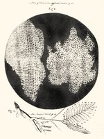
Robert Hooke (1665): English scientist who first observed and named "cells" in cork using a primitive microscope. His drawings illustrated the compartmentalized structure of cork tissue.

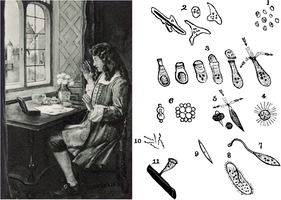
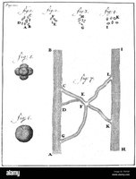
Antonie van Leeuwenhoek (1674): Dutch scientist, known as the father of biological microscopy, observed single-celled organisms ("animalcules"), bacteria, yeast, blood cells, and capillaries in water and other samples.

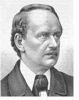
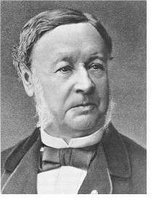
Cell Theory (1839): Formulated by M.J. Schleiden and Theodor Schwann, stating that all organisms are composed of cells and that cells are the fundamental unit of life.
Characteristics and Types of Cells
Universal Features of Cells
All cells share certain structural and functional characteristics:
Plasma Membrane: Encloses the cell, acting as a selective barrier.
Cytosol: Fluid interior where cellular processes occur.
Genetic Material: DNA (and RNA) for inheritance and function.
Energy Transformation: Cells can transform and use energy.
Prokaryotic Cells
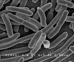
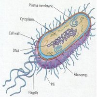
Prokaryotes are simpler, smaller cells lacking a nucleus and membrane-bound organelles. Their DNA is located in a nucleoid region and is typically circular.
Size: 1–5 µm
Structure: No nucleus, no organelles, unicellular
Examples: Bacteria and Archaea
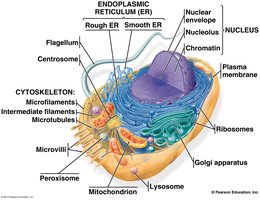
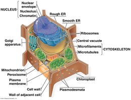
Eukaryotic Cells
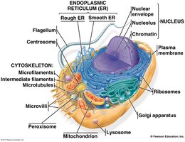
Eukaryotes have a nucleus and various membrane-bound organelles. They can be unicellular or multicellular and are generally larger than prokaryotes.
Size: 5–100 µm
Structure: Nucleus, linear chromosomes, many organelles
Examples: Animals, plants, fungi, protists
Cell Size and Surface Area
Why Cells Are Small
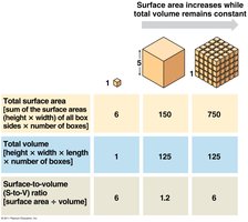
Cell size is limited by the surface area-to-volume ratio, which affects the efficiency of material exchange and internal communication.
Higher Surface/Volume Ratio: Facilitates import/export of nutrients and wastes.
Rapid Internal Exchange: Components are close for efficient interaction.
Cube Size | Surface Area | Volume | Surface/Volume Ratio |
|---|---|---|---|
1 | 6 | 1 | 6 |
5 | 150 | 125 | 1.2 |
Many small cubes | 750 | 125 | 6 |
Cell Shapes and Structure-Function Relationship
Cell Shapes
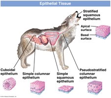
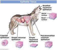
Cell shape is closely related to function. Common shapes include squamous (flat), columnar (columns), cuboidal (cubes), and spherical.
Squamous: Flat cells, often found in lining tissues.
Columnar: Tall, column-like cells, specialized for absorption.
Cuboidal: Cube-shaped cells, often involved in secretion.
Spherical: Round cells, such as some blood cells.
Molecular Components of Cells
Major Biomolecules
Cells are composed of water, proteins, lipids, carbohydrates, and nucleic acids. Their proportions vary, but water is the most abundant.
Water: ~80%
Protein: ~15%
Lipid: ~3%
Carbohydrates: ~1%
Nucleic Acids: ~1%
Cell Membrane Structure and Function
Phospholipid Bilayer
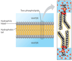
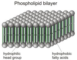
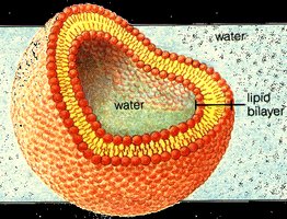
The cell membrane is primarily composed of a phospholipid bilayer, which forms a selective barrier between the cell and its environment.
Phospholipids: Amphipathic molecules with hydrophilic heads and hydrophobic tails.
Membrane Structure: Sandwich-like arrangement, with hydrophobic tails facing inward and hydrophilic heads facing outward.
Tail Saturation and Membrane Fluidity
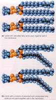
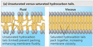
The degree of saturation of phospholipid tails affects membrane fluidity.
Saturated Tails: Pack tightly, making the membrane more viscous.
Unsaturated Tails: Kinked, prevent tight packing, increase fluidity.
Cholesterol
Cholesterol is interspersed within the membrane, modulating fluidity and stability.
Function: Prevents extremes of fluidity and rigidity.
Membrane Proteins
Proteins embedded in the membrane perform essential functions such as transport, communication, and cell-cell connections.
Integral (Transmembrane) Proteins: Span the membrane, involved in transport.
Peripheral Proteins: Attached to one side, involved in signaling and structure.
Hydrophobic/Polar Regions: Internal regions are hydrophobic; external regions are hydrophilic.
Fluid-Mosaic Model
The cell membrane is described by the fluid-mosaic model, where lipids and proteins move laterally within the bilayer, creating a dynamic structure.
Carbohydrates in Membranes
Carbohydrates attached to proteins (glycoproteins) or lipids (glycolipids) are important for cellular signaling and identification.
Membrane Function: Movement of Substances
Selective Permeability
The plasma membrane is selectively permeable, allowing certain substances to pass while blocking others.
Passive Movement
Passive transport does not require energy and occurs down a concentration gradient.
Simple Diffusion: Random movement of small, nonpolar molecules, gases, and lipids through the membrane.
Channel-Mediated Diffusion: Transmembrane proteins form channels for selective passage of ions and large polar molecules.
Carrier-Mediated Diffusion: Carrier proteins transport specific molecules across the membrane.
Osmosis
Osmosis is the diffusion of water across a semi-permeable membrane from higher to lower concentration.
Isosmotic (Isotonic): Equal solute and solvent concentrations; no net water movement.
Hyposmotic (Hypotonic): Lower solute concentration; water moves away.
Hyperosmotic (Hypertonic): Higher solute concentration; water moves in.
Active Movement
Active transport requires energy (ATP) to move substances against their concentration gradient.
Active Transport: Carrier proteins (pumps) move molecules from low to high concentration.
Cotransport: Two or more molecules are transported together, often using gradients established by pumps.
Sodium-Potassium Pump: Maintains electrochemical gradients in animal cells.
Bulk Movement
Bulk movement involves the transport of large quantities or objects via vesicles.
Endocytosis: Bringing materials into the cell; includes phagocytosis (cellular eating) and pinocytosis (cellular drinking).
Exocytosis: Secretion of materials by fusion of vesicles with the plasma membrane.
Plant Cell Walls
Structure and Function
Plant cell walls provide shape, strength, and protection. They are composed of cellulose, pectins, lignins, and lipids.
Cellulose: Most abundant organic molecule, forms a strong matrix.
Pectins: Hydrophilic polysaccharides, create a jelly-like matrix.
Lignins: Ringed molecules, harden and strengthen the wall.
Lipids: Form a waxy cuticle, reduce water loss.
Wall Layers
Primary Cell Wall: Thin, flexible, formed during cell growth.
Secondary Cell Wall: Tougher, more water-resistant, formed as cell matures.
Middle Lamella: Pectin-rich layer, glues cells together.
Summary Table: Prokaryotic vs. Eukaryotic Cells
Feature | Prokaryotes | Eukaryotes |
|---|---|---|
Nucleus | No | Yes |
DNA Form | Circular | Linear |
Organelles | No | Yes |
Size | 1–5 µm | 5–100 µm |
Examples | Bacteria, Archaea | Animals, Plants, Fungi, Protists |
