 Indietro
Cell Structure, Membrane Function, and Molecular Genetics: Study Notes for General Biology
Guida di studio - Note intelligenti
 Appunti personalizzati basati sui tuoi materiali, ampliati con definizioni chiave, esempi e contesto.
Cell Structure and Function
Overview of Cell Types
Cells are the fundamental units of life, and all living organisms are composed of cells. There are two main types of cells: prokaryotic and eukaryotic. Both share basic features such as a plasma membrane, cytoplasm, chromosomes, and ribosomes, but differ in complexity and internal organization.
Prokaryotic cells: Lack a nucleus; genetic material is found in the nucleoid region.
Eukaryotic cells: Have a nucleus surrounded by a nuclear envelope; contain membrane-bound organelles.
Surface area to volume ratio is a key factor limiting cell size; smaller cells have a higher ratio, facilitating efficient exchange of materials.
Major Cell Organelles and Their Functions
Each organelle within a eukaryotic cell has a specific function, contributing to the cell's overall operation. The table below summarizes key organelles, their functions, and common analogies.
Organelle/Part of Cell | Functions | Analogy |
|---|---|---|
Nucleus | Stores genetic information; controls cell activities | Control center |
Nucleolus | Produces ribosomes | Factory assembly line |
Cytoplasm | Site of metabolic reactions; suspends organelles | Factory floor |
Rough ER | Protein synthesis and processing | Production line |
Smooth ER | Lipid synthesis; detoxification | Oil refinery |
Ribosomes | Protein synthesis | Machines |
Golgi apparatus | Modifies, sorts, and packages proteins/lipids | Shipping center |
Vesicles | Transport materials within cell | Delivery trucks |
Lysosomes | Digest waste and cellular debris | Recycling center |
Peroxisomes | Break down fatty acids and detoxify | Hazardous waste facility |
Mitochondria | Produce ATP via cellular respiration | Power plant |
Cell membrane | Regulates entry/exit of substances | Security gate |
Cellular Diseases and Organelle Dysfunction
Malfunction of specific organelles can lead to various diseases. The table below highlights examples:
Disease or Syndrome | Symptoms | Organelle Normal Function | Affected Organelle |
|---|---|---|---|
Cystic Fibrosis | Thick mucus, lung infections, digestive issues | Regulates movement of materials in/out of cell | Cell membrane |
ALD | Fatty acid buildup, lethargy, loss of limb control | Break down fatty acids and toxic cell products | Peroxisomes |
Pompé | Weakness, "floppy" appearance, breathing difficulties | Break down macromolecules | Lysosomes |
Kartagener | Breathing problems, sinus/ear infections, infertility | Movement of materials in lungs and fallopian tubes | Cilia/flagella |
Membrane Structure and Function
Fluid Mosaic Model
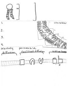
The fluid mosaic model describes the structure of the plasma membrane as a dynamic combination of lipids and proteins. The membrane is composed of a phospholipid bilayer with embedded proteins, cholesterol, glycoproteins, and glycolipids.
Phospholipids: Amphipathic molecules with hydrophilic heads and hydrophobic tails.
Proteins: Integral and peripheral proteins serve functions such as transport, enzymatic activity, and cell signaling.
Cholesterol: Modulates membrane fluidity.
Membrane asymmetry: Different composition on inner and outer leaflets.
Unsaturated fatty acids increase fluidity, while saturated fatty acids decrease it.
Membrane Transport Mechanisms
The plasma membrane exhibits selective permeability, allowing certain substances to cross while blocking others. Transport occurs via:
Diffusion: Passive movement of molecules from high to low concentration.
Facilitated diffusion: Passive transport via carrier or channel proteins.
Active transport: Movement against concentration gradient, requiring energy (ATP).
Osmosis: Diffusion of water across a membrane.
Endocytosis/Exocytosis: Bulk transport of materials into/out of cell.
Key terms: hypertonic (higher solute), hypotonic (lower solute), isotonic (equal solute).
Molecular Genetics: DNA and RNA
Structure of DNA and RNA
DNA is a double-stranded, antiparallel molecule composed of nucleotides (phosphate, deoxyribose, nitrogenous base). RNA is single-stranded and contains uracil instead of thymine.
Purines: Adenine (A), Guanine (G)
Pyrimidines: Cytosine (C), Thymine (T), Uracil (U)
Phosphodiester bonds: Link nucleotides in a strand
Antiparallel: Strands run in opposite directions (5' to 3' and 3' to 5')
DNA Replication
DNA replication is the process by which a cell copies its DNA before cell division. It is semi-conservative, meaning each new DNA molecule contains one old and one new strand.
Key enzymes: Helicase (unwinds DNA), Primase (adds RNA primer), DNA polymerase III (synthesizes new strand), DNA polymerase I (removes primer), Ligase (joins fragments)
Leading strand: Synthesized continuously
Lagging strand: Synthesized in Okazaki fragments
Replication fork: Site of active DNA synthesis
Experiments by Meselson and Stahl confirmed the semi-conservative model.
Gene Expression: Transcription and Translation
Gene expression involves two main steps: transcription (DNA to mRNA) and translation (mRNA to protein).
Transcription: RNA polymerase binds to promoter, synthesizes mRNA from DNA template.
RNA processing: Addition of 5' cap, poly-A tail, and splicing out introns.
Translation: Ribosomes read mRNA codons, tRNA brings amino acids, rRNA catalyzes peptide bond formation.
Genetic code: Triplet codons specify amino acids; start codon (AUG), stop codons (UAA, UAG, UGA).
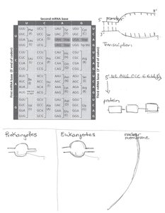
Prokaryotes can transcribe and translate simultaneously, while eukaryotes separate these processes by the nuclear membrane.
Microscopy and Cell Visualization
Types of Microscopes
Microscopes are essential tools for studying cells. The main types include:
Light microscope (LM): Uses visible light; allows observation of living cells.
Electron microscope (EM): Uses electron beams; higher resolution.
Scanning electron microscope (SEM): Surface details.
Transmission electron microscope (TEM): Internal structures.
Key concepts: magnification (enlargement), resolution (clarity), contrast (distinguishing features).
Cellular Connections and Structures
Cell Junctions and Extracellular Matrix
Cells are connected and supported by various structures:
Tight junctions: Prevent leakage between cells.
Desmosomes: Anchor cells together.
Gap junctions: Allow communication between cells.
Extracellular matrix (ECM): Provides structural support outside the cell.
Plasmodesmata: Channels between plant cells.
These structures are essential for tissue integrity and intercellular communication.
Cytoskeleton and Cell Movement
Cytoskeletal Elements
The cytoskeleton is a network of protein filaments that provides structural support and enables movement.
Microtubules: Hollow tubes; involved in cell shape, transport, and division.
Microfilaments: Actin filaments; support cell shape and movement.
Intermediate filaments: Provide mechanical strength.
Motor proteins: Move along cytoskeletal tracks (e.g., myosin, kinesin, dynein).
Flagella and cilia: Structures for cell movement.
These elements are crucial for processes such as intracellular transport, cell division, and motility.
