 Indietro
Cell Structure, Membrane Function, and Prokaryotic Adaptations: Study Notes for General Biology
Guida di studio - Note intelligenti
 Appunti personalizzati basati sui tuoi materiali, ampliati con definizioni chiave, esempi e contesto.
Cell Structure and Compartmentalization
Overview of Cell Theory
Cell theory is a fundamental concept in biology, stating that all living things are composed of cells, cells are the basic unit of life, and all cells arise from pre-existing cells. Cells exhibit compartmentalization, which allows for specialized functions within distinct organelles.
Compartmentalization: Organelles are membrane-bound structures that create micro-environments for optimal biochemical reactions.
Cell Types: Cells are classified as prokaryotic (lacking membrane-bound organelles) or eukaryotic (containing membrane-bound organelles).
Minimum Requirements: All cells possess a plasma membrane, genetic material (DNA), and cytoplasm with ribosomes.
Cell Size and Microscopy
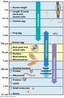
Cells vary greatly in size, from microscopic bacteria to larger plant and animal cells. The size of cells is limited by the surface area-to-volume ratio, which affects the efficiency of material exchange.
Surface Area-to-Volume Ratio: As cells increase in size, their volume grows faster than their surface area, limiting the rate of diffusion and transport.
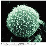
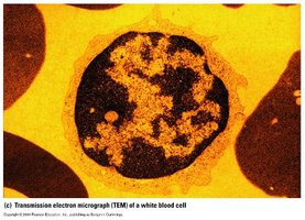
Microscopy: Different types of microscopy (light, electron) are used to visualize cells and their components.
Prokaryotic vs. Eukaryotic Cells
Prokaryotic cells lack membrane-bound organelles and are generally smaller, while eukaryotic cells have complex internal structures.
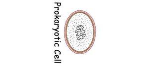
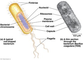
Prokaryotic Cell: Contains a plasma membrane, DNA in a nucleoid region, ribosomes, and cytoplasm.
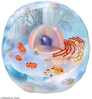
Eukaryotic Cell: Contains a nucleus, endoplasmic reticulum, Golgi apparatus, mitochondria, and other organelles.
Membrane Structure and Function
Phospholipid Bilayer and Fluid Mosaic Model
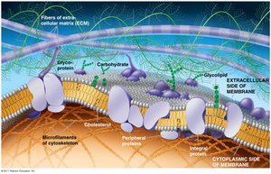
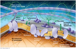
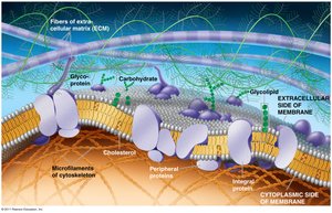
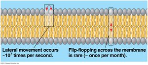
The plasma membrane is composed of a phospholipid bilayer with embedded proteins, forming a selectively permeable barrier. The fluid mosaic model describes the dynamic nature of the membrane.
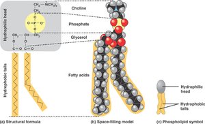
Phospholipids: Amphipathic molecules with hydrophilic heads and hydrophobic tails, spontaneously forming bilayers in aqueous environments.
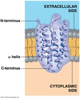
Proteins: Integral and peripheral proteins serve functions such as transport, enzymatic activity, signal transduction, and cell recognition.
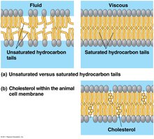
Fluidity: Membrane fluidity is influenced by fatty acid composition and cholesterol content.
Membrane Transport Mechanisms
Cell membranes regulate the movement of substances through passive and active transport mechanisms, maintaining homeostasis.
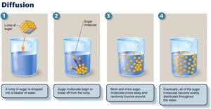
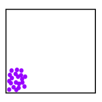
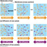
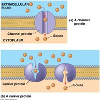
Passive Transport: Includes diffusion and facilitated diffusion, where molecules move down their concentration gradient without energy input.
Active Transport: Requires energy (usually ATP) to move molecules against their concentration gradient.
Selective Permeability: The membrane allows certain molecules (e.g., O2, CO2) to pass freely, while others require transport proteins.
Osmosis and Biological Consequences
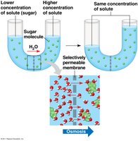
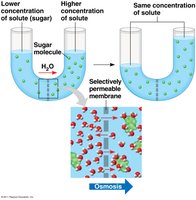
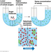
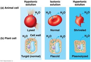
Osmosis is the diffusion of water across a selectively permeable membrane. The osmotic balance affects cell shape and function, especially in plant and animal cells.
Hypotonic Solution: Water enters the cell, causing it to swell (turgid in plants, lysed in animals).
Isotonic Solution: No net movement of water; cell remains normal.
Hypertonic Solution: Water leaves the cell, causing it to shrink (plasmolyzed in plants, shriveled in animals).
Turgor Pressure: In plant cells, osmotic pressure creates turgor, essential for structural support.
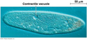
Contractile Vacuoles: Some protists use contractile vacuoles to expel excess water.
Bulk Transport: Endocytosis and Exocytosis
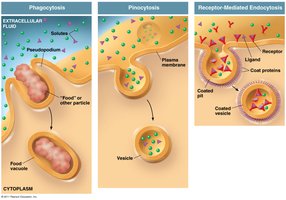
Cells move large quantities of material in and out through endocytosis and exocytosis, processes that involve vesicle formation and fusion with the membrane.
Endocytosis: The cell engulfs material, forming vesicles for transport.
Exocytosis: Vesicles fuse with the plasma membrane to release contents outside the cell.
Organelles and Their Functions
Nucleus and Ribosomes
The nucleus is the control center of the cell, containing genetic material. Ribosomes are the sites of protein synthesis.
Nucleus: Enclosed by a double membrane; nucleolus is the site of ribosomal RNA synthesis.
Ribosomes: Can be free in the cytoplasm or attached to the endoplasmic reticulum; number varies by cell type.
Endoplasmic Reticulum and Golgi Apparatus
The endoplasmic reticulum (ER) is involved in synthesis and transport. The Golgi apparatus modifies, sorts, and packages proteins and lipids.
Smooth ER: Lipid synthesis, carbohydrate metabolism, detoxification, calcium storage.
Rough ER: Protein synthesis (with ribosomes), membrane production.
Golgi Apparatus: Modifies molecules, attaches "tags" for vesicle targeting.
Lysosomes, Vacuoles, and Peroxisomes
Lysosomes digest cellular waste, vacuoles store substances, and peroxisomes detoxify harmful compounds.
Lysosomes: Contain digestive enzymes; produced in the ER and processed in the Golgi.
Vacuoles: Central vacuole in plants stores water, ions, pigments, toxins; contractile vacuole in protists expels water.
Peroxisomes: Carry out oxidation reactions, convert hydrogen peroxide to water.
Chloroplasts and Mitochondria
Chloroplasts and mitochondria are energy-converting organelles. Chloroplasts perform photosynthesis; mitochondria carry out cellular respiration.
Chloroplasts: Convert light energy to chemical energy (glucose).
Mitochondria: Harvest energy from food molecules to produce ATP.
Cytoskeleton
The cytoskeleton provides structural support, organization, and movement within the cell.
Microtubules: Hollow tubes involved in cell shape, transport, and division.
Microfilaments: Actin filaments involved in cell movement and shape.
Intermediate Filaments: Provide mechanical support.
Prokaryotic Cell Structure and Adaptations
Prokaryotic Diversity and Adaptation
Prokaryotes are highly adaptable and thrive in extreme environments. They are divided into two domains: Bacteria and Archaea.
Size: Prokaryotic cells are typically 0.5–5 µm, much smaller than eukaryotic cells.
Shapes: Common shapes include cocci (spherical), bacilli (rod-shaped), and spirals.
Cell-Surface Structures
Prokaryotic cells possess unique surface structures that contribute to their survival and adaptation.
Cell Wall: Maintains shape, protects, and prevents bursting. Bacterial cell walls contain peptidoglycan; archaea lack peptidoglycan.
Gram Stain: Used to classify bacteria based on cell wall composition (Gram-positive vs. Gram-negative).
Capsule: Polysaccharide or protein layer that protects and aids in attachment.
Fimbriae and Pili: Structures for attachment and DNA exchange.
Endospores: Metabolically inactive structures for survival in harsh conditions.
Motility and Flagella
Many prokaryotes are motile, using flagella to move toward or away from stimuli (taxis).
Flagella: Structures for movement; differ in composition between bacteria, archaea, and eukaryotes.
Chemotaxis: Movement in response to chemical signals.
Summary Table: Prokaryotic vs. Eukaryotic Cells
Feature | Prokaryotic Cell | Eukaryotic Cell |
|---|---|---|
Size | 0.5–5 µm | 10–100 µm |
Nucleus | Absent | Present |
Organelles | Absent | Present |
Cell Wall | Peptidoglycan (Bacteria) | Cellulose (Plants), Chitin (Fungi) |
DNA Location | Nucleoid | Nucleus |
Division | Binary Fission | Mitosis/Meiosis |
Additional info: Some details and examples were inferred for completeness and clarity, such as the summary table and expanded explanations of cell transport and organelle functions.
