 Indietro
Cell Structure, Membrane Transport, and Cell Signaling: Study Notes for General Biology
Guida di studio - Note intelligenti
 Appunti personalizzati basati sui tuoi materiali, ampliati con definizioni chiave, esempi e contesto.
Cell Structure and Function
Microscopy and Cell Fractionation
Biologists use microscopy and biochemistry to study cells. Improvements in magnification, resolution, and contrast have advanced our understanding of cell structure. Light microscopy (LM) allows observation of living cells, while electron microscopy (EM) provides higher resolution for detailed structures. Cell fractionation uses centrifugation to separate cellular components for biochemical analysis.
Magnification: Enlarges the image of the specimen.
Resolution: Distinguishes fine details and structures.
Contrast: Enhances differences between parts of the sample.
Cell fractionation: Sequential centrifugation isolates organelles.
Example: Studying mitochondria by isolating them from cell lysates.
Prokaryotic vs. Eukaryotic Cells
All cells are bounded by a plasma membrane. Prokaryotic cells lack nuclei and membrane-bound organelles, while eukaryotic cells have internal membranes that compartmentalize functions. The surface-to-volume ratio affects cell size and efficiency.
Prokaryotes: No nucleus, no membrane-bound organelles.
Eukaryotes: Nucleus, endoplasmic reticulum, Golgi apparatus, mitochondria, and (in plants) chloroplasts.
Surface-to-volume ratio: Smaller cells have higher ratios, facilitating exchange of materials.
Example: Plant cells contain chloroplasts for photosynthesis; animal cells do not.
Cell Components and Their Functions
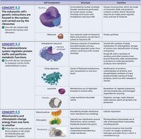
Major organelles and their functions are summarized below:
Cell Component | Structure | Function |
|---|---|---|
Nucleus | Enclosed by nuclear envelope; contains nucleolus and chromatin | Genetic information storage; ribosome synthesis |
Ribosome | Complex of RNA and protein | Protein synthesis |
Endoplasmic Reticulum (ER) | Network of membranous tubules | Rough ER: protein synthesis; Smooth ER: lipid synthesis |
Golgi Apparatus | Stack of flattened membranes | Modification, sorting, and packaging of proteins and lipids |
Lysosome | Membrane-bound sac | Digestion of macromolecules |
Mitochondrion | Double membrane, inner folds (cristae) | ATP production (cellular respiration) |
Chloroplast | Double membrane, internal thylakoids | Photosynthesis (in plants) |
Membrane Structure and Function
Fluid Mosaic Model
The fluid mosaic model describes the plasma membrane as a dynamic structure with proteins embedded in a phospholipid bilayer. Phospholipids have hydrophilic heads and hydrophobic tails, allowing lateral movement and flexibility. Cholesterol and unsaturated fatty acids influence membrane fluidity.
Amphipathic proteins: Embedded in the bilayer, perform various functions.
Membrane proteins: Transport, enzymatic activity, cell-cell recognition, signal transduction.
Glycoproteins and glycolipids: Short sugar chains on the membrane exterior, involved in cell recognition.
Selective Permeability
The plasma membrane is selectively permeable, allowing certain molecules to pass while restricting others. Hydrophobic molecules pass easily; polar molecules and ions require transport proteins.
Hydrophobic molecules: Soluble in lipids, diffuse rapidly.
Transport proteins: Facilitate movement of polar molecules and ions.
Example: Aquaporins increase water permeability.
Passive Transport: Diffusion and Facilitated Diffusion
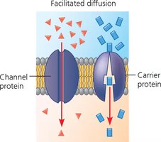
Passive transport is the movement of substances down their concentration gradient without energy input. Facilitated diffusion uses channel and carrier proteins to transport molecules across the membrane.
Diffusion: Spontaneous movement from high to low concentration.
Facilitated diffusion: Channel proteins form pores; carrier proteins change shape to move solutes.
Osmosis: Diffusion of water across a membrane.
Active Transport
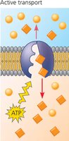
Active transport moves solutes against their concentration gradient using energy, usually from ATP. Membrane proteins (pumps) are required for this process.
ATP: Provides energy for transport.
Electrochemical gradient: Combination of concentration and electrical gradients.
Cotransport: One solute's downhill diffusion drives another's uphill transport.
Bulk Transport: Endocytosis and Exocytosis
Cells use endocytosis and exocytosis for bulk transport of large molecules. Endocytosis includes phagocytosis, pinocytosis, and receptor-mediated endocytosis.
Phagocytosis: Cell engulfs large particles.
Pinocytosis: Cell takes in fluid and small solutes.
Receptor-mediated endocytosis: Specific molecules bind to membrane receptors for uptake.
Cell Signaling
Cell Signaling Pathways
Cells communicate via signaling molecules that bind to receptors, triggering a cascade of events leading to a cellular response. The process involves three stages: reception, transduction, and response.
Reception: Signaling molecule (ligand) binds to receptor protein.
Transduction: Relay molecules transmit the signal, often via phosphorylation cascades.
Response: Activation of cellular processes, such as gene expression or enzyme activity.
Types of Signaling
Local signaling: Direct contact or secretion of growth factors.
Long-distance signaling: Hormones travel through the bloodstream (animals) or sap (plants).
Membrane receptors: G protein-coupled receptors (GPCRs), ligand-gated ion channels.
Intracellular receptors: For hydrophobic signaling molecules (e.g., steroid hormones).
Signal Transduction Mechanisms
Phosphorylation cascades: Series of protein kinases activate each other by adding phosphate groups.
Second messengers: Small molecules like cyclic AMP (cAMP) amplify and broadcast signals.
Protein phosphatases: Remove phosphate groups, regulating pathway activity.
Cellular Responses
Regulation of transcription: Activation or repression of gene expression in the nucleus.
Regulation of cytoplasmic activities: Changes in enzyme activity, cytoskeleton, or metabolism.
Example: Epinephrine triggers glycogen breakdown in liver cells.
Summary Table: Membrane Transport Mechanisms
Transport Type | Energy Required | Direction | Example |
|---|---|---|---|
Simple Diffusion | No | Down gradient | O2, CO2 |
Facilitated Diffusion | No | Down gradient | Glucose via carrier protein |
Active Transport | Yes (ATP) | Against gradient | Na+/K+ pump |
Bulk Transport | Yes | Variable | Endocytosis, exocytosis |
Additional info: These notes cover key concepts from General Biology chapters on cell structure, membrane transport, and cell signaling, including definitions, examples, and mechanisms. The included images directly illustrate facilitated diffusion, active transport, cell component functions, and cell signaling pathways, reinforcing the explanations provided.
