 Indietro
Cell Structure, Organelles, Cytoskeleton, Cell Junctions, and Energy Metabolism: General Biology Study Notes
Guida di studio - Note intelligenti
 Appunti personalizzati basati sui tuoi materiali, ampliati con definizioni chiave, esempi e contesto.
Inside the Cell: Structure and Function
Prokaryotic vs. Eukaryotic Cells
Cells are the fundamental units of life, classified as prokaryotic or eukaryotic based on their structural features.
Prokaryotes: Lack a membrane-bound nucleus; include Bacteria and Archaea. Typically small and unicellular, with a plasma membrane, tough cell wall (peptidoglycan in bacteria), ribosomes, DNA, plasmids, and sometimes flagella. DNA is supercoiled to fit inside the cell.
Eukaryotes: Possess a nucleus and extensive internal membrane systems. Usually larger, with diverse organelles and a dynamic cytoskeleton.
Cytoplasm refers to all contents within the cell, excluding the nucleus in eukaryotes.
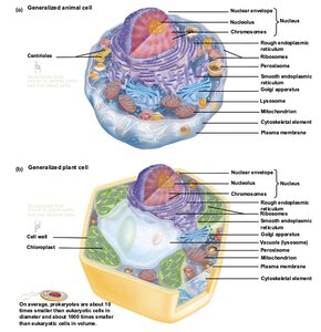
Major Differences Between Eukaryotes and Prokaryotes
Eukaryotic chromosomes are enclosed in a nucleus.
Eukaryotic cells are larger and contain more internal membranes.
Eukaryotic cells have a dynamic cytoskeleton.
Eukaryotic Organelles and Their Functions
Overview of Organelles
Eukaryotic cells contain specialized, membrane-bound organelles that compartmentalize cellular functions, increasing efficiency and allowing for complex processes.
Nucleus: Stores genetic information, assembles ribosomal subunits, and provides structural support. Surrounded by a double membrane (nuclear envelope) with nuclear pores.
Ribosomes: Sites of protein synthesis; consist of large and small subunits made of RNA and protein. Not membrane-bound.
Endoplasmic Reticulum (ER):
Rough ER: Studded with ribosomes; synthesizes and processes proteins.
Smooth ER: Lacks ribosomes; synthesizes lipids and stores Ca2+.
Golgi Apparatus: Processes, sorts, and ships proteins; consists of stacked cisternae.
Peroxisomes: Catalyze oxidation reactions, break down fatty acids.
Lysosomes: Digest and recycle cellular waste; contain acid hydrolases.
Vacuoles: Storage and digestion in plants and fungi; maintain cell volume.
Mitochondria: Site of ATP production; contain their own DNA and ribosomes.
Chloroplasts: Conduct photosynthesis in plants and algae; contain thylakoids and their own DNA.
Cell Wall: Provides structural support; composed of cellulose (plants/algae) or chitin (fungi).
Cytoskeleton: Provides structural support, cell shape, and facilitates movement.
Plasma Membrane: Maintains selective permeability and intracellular environment.
Lysosome Function and Recycling Pathways
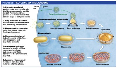
Lysosomes are central to cellular digestion and recycling, utilizing three main pathways: phagocytosis, autophagy, and receptor-mediated endocytosis.
Phagocytosis: Engulfment of foreign particles by phagocytes, forming phagosomes that fuse with lysosomes.
Autophagy: Degradation of damaged organelles or cytoplasmic components via autophagosomes.
Receptor-mediated endocytosis: Uptake of specific molecules via vesicles, which mature into lysosomes.
Protein Trafficking and the Endomembrane System
Protein Sorting and Signal Hypothesis
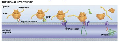
Proteins are directed to their correct cellular locations by signal sequences, which are recognized during translation.
Proteins destined for the endomembrane system possess an ER signal sequence, recognized by the signal recognition particle (SRP).
SRP binds to the SRP receptor on the ER membrane, allowing the protein to enter the ER.
Secretory Pathway
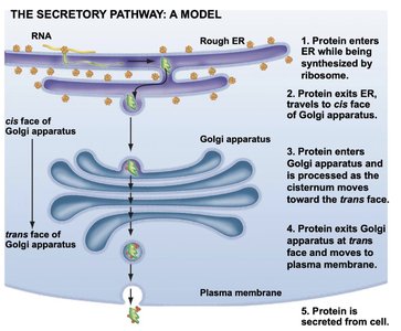
The secretory pathway describes the journey of proteins from synthesis to secretion.
Proteins enter the ER during synthesis.
They are transported to the Golgi apparatus for processing.
Processed proteins are sorted and shipped to their final destinations, including secretion from the cell.
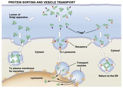
Protein Sorting and Vesicle Transport
Proteins are tagged and sorted in the Golgi apparatus, then transported in vesicles to their destinations.
Tags (such as phosphorylated sugars) direct proteins to lysosomes or other organelles.
Transport vesicles interact with receptors to deliver contents.
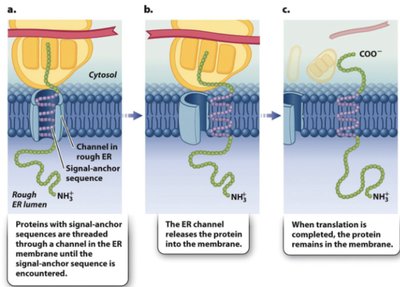
Transmembrane Protein Targeting
Transmembrane proteins are inserted into the ER membrane during translation, guided by signal-anchor sequences.
Signal-anchor sequence is recognized and threaded through the ER channel.
Protein remains embedded in the membrane after translation.
The Cytoskeleton: Structure and Function
Types of Cytoskeletal Elements
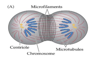
The cytoskeleton is a dynamic network of protein fibers that provides structural support, facilitates movement, and organizes cellular components.
Actin Filaments (Microfilaments): Smallest, double helix of actin monomers; involved in cell shape, movement, and muscle contraction.
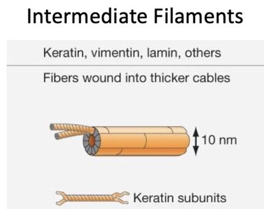
Intermediate Filaments: Medium-sized, composed of various proteins (e.g., keratin, vimentin, lamin); provide structural support.
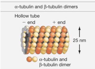
Microtubules: Largest, hollow tubes of α- and β-tubulin dimers; involved in cell shape, division, and intracellular transport.
Microtubule-Based Transport and Motility
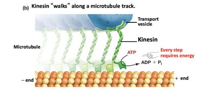
Microtubules serve as tracks for motor proteins such as kinesin, which transport vesicles and organelles within the cell.
Kinesin "walks" along microtubules, powered by ATP hydrolysis.
Microtubules also form the structural basis for cilia and flagella, which enable cell movement.
Cilia, Flagella, and the Axoneme
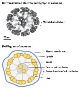
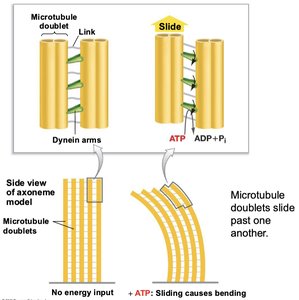
Cilia and flagella are motile structures composed of microtubules arranged in a 9+2 pattern (axoneme), associated with dynein motor proteins.
Dynein arms cause sliding of microtubule doublets, resulting in bending and movement.
Actin-Myosin Interactions and Muscle Contraction
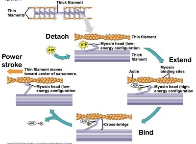
Actin filaments interact with myosin motor proteins to produce movement, including muscle contraction.
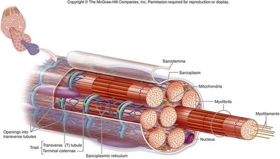
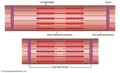
Muscle fibers contain myofibrils composed of actin (thin) and myosin (thick) filaments.
Contraction occurs via the sliding filament model, where myosin heads bind to actin and pull filaments toward the center of the sarcomere.
Cell Junctions and the Extracellular Matrix
Cell Junctions: Types and Functions
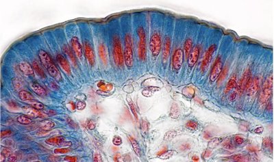
Cell junctions are specialized structures that connect cells to each other and to the extracellular matrix, providing adhesion, communication, and barrier functions.
Anchoring Junctions:
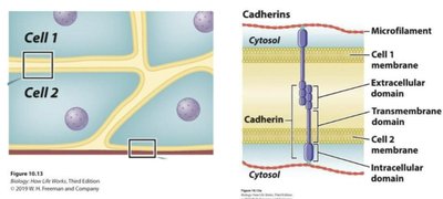
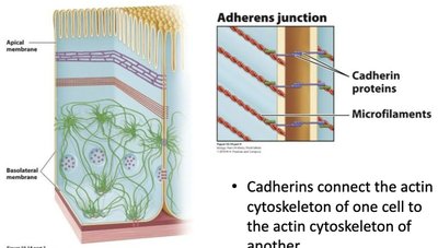
Adherens Junctions: Cadherins connect actin cytoskeletons of adjacent cells.
Desmosomes: Cadherins connect intermediate filaments for strong adhesion.
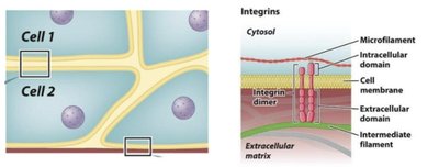
Hemidesmosomes: Integrins connect intermediate filaments to the extracellular matrix.
Barrier Junctions:
Tight Junctions: Claudins and occludins create a seal, preventing leakage between cells.
Communicating Junctions:
Gap Junctions: Connexins form channels for ion and molecule exchange between animal cells.
Plasmodesmata: Channels between plant cells for communication.
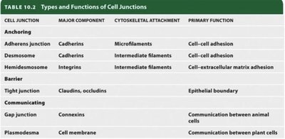
Cell Junction | Major Component | Cytoskeletal Attachment | Primary Function |
|---|---|---|---|
Adherens Junction | Cadherins | Microfilaments | Cell–cell adhesion |
Desmosome | Cadherins | Intermediate filaments | Cell–cell adhesion |
Hemidesmosome | Integrins | Intermediate filaments | Cell–extracellular matrix adhesion |
Tight Junction | Claudins, occludins | None | Epithelial boundary |
Gap Junction | Connexins | None | Communication between animal cells |
Plasmodesma | Cell membrane | None | Communication between plant cells |
Extracellular Matrix (ECM)
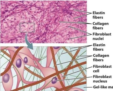
The ECM is a meshwork of proteins and polysaccharides that provides structural support, cell shape, and signaling in animal tissues.
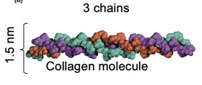
Collagen: Most abundant protein in animals; provides tensile strength via triple helix structure and fiber formation.
Elastin: Provides flexibility; composed of cross-linked, unstructured polypeptide chains.
Energy Metabolism and Enzymes
Metabolism: Catabolism and Anabolism
Metabolism encompasses all biochemical reactions in the cell, divided into catabolic (breakdown, energy release) and anabolic (building, energy consumption) pathways.
Catabolism: Breakdown of molecules; releases energy, often as ATP.
Anabolism: Synthesis of molecules; requires energy input.
Energy Types and Thermodynamics
Cells utilize kinetic and potential energy, governed by the laws of thermodynamics.
Kinetic Energy: Energy of motion (thermal, light, electrical).
Chemical Potential Energy: Stored in molecular bonds.
First Law: Energy is conserved; cannot be created or destroyed.
Second Law: Energy transformations increase entropy (disorder).
Gibbs Free Energy and Reaction Spontaneity
The spontaneity of a reaction is determined by the change in Gibbs free energy ().
: Spontaneous, exergonic
: Nonspontaneous, endergonic
ATP and Energetic Coupling
ATP hydrolysis is highly exergonic and is used to drive endergonic reactions via energetic coupling.
Hydrolysis of ATP releases energy and increases entropy.
Coupling occurs via phosphorylation of substrates.
Enzymes: Catalysis and Regulation
Enzymes are biological catalysts that lower activation energy and increase reaction rates without altering .
Enzyme-substrate binding occurs at the active site, often via induced fit.
Enzyme action involves initiation, transition state facilitation, and termination.
Regulation includes competitive inhibition, allosteric regulation, and negative feedback.
Cellular Respiration
Overview and Steps
Cellular respiration is a catabolic, exergonic process that oxidizes glucose to CO2 and H2O, generating ATP through a series of redox reactions.
Overall reaction:
Redox reactions: Oxidation (loss of electrons), reduction (gain of electrons).
Electron carriers: NAD+ and FAD are reduced to NADH and FADH2.
Stages of Cellular Respiration
Glycolysis: Occurs in cytosol; breaks down glucose to pyruvate, yields ATP and NADH.
Pyruvate Processing: Pyruvate is oxidized to acetyl CoA in mitochondria, producing NADH and CO2.
Citric Acid Cycle: Acetyl CoA is oxidized to CO2, yielding NADH, FADH2, GTP/ATP.
Electron Transport Chain and Chemiosmosis: Reduced carriers are oxidized, driving ATP synthesis.
Regulation: Feedback inhibition (e.g., ATP inhibits phosphofructokinase) ensures efficient energy use.
*Additional info: Some details and explanations were expanded for clarity and completeness, including definitions, examples, and context for key terms and processes. Tables and images were included only when directly relevant to the adjacent content, as per instructions.*
