 Indietro
Cell Structures and Functions: General Biology Study Guide
Guida di studio - Note intelligenti
 Appunti personalizzati basati sui tuoi materiali, ampliati con definizioni chiave, esempi e contesto.
Cell Theory and Studying Cells
Historical Foundations of Cell Theory
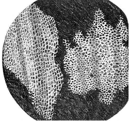
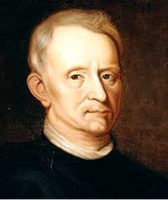
The cell theory is a fundamental concept in biology, describing the properties and significance of cells. Early scientists such as Robert Hooke and Anton van Leeuwenhoek contributed to the discovery and understanding of cells.
Robert Hooke: First described cells in cork using a microscope.
Anton van Leeuwenhoek: Observed living cells, referred to as "animalcules."
Schleiden and Schwann: Formulated the cell theory.
Cell Theory:
All living things are composed of cells.
Cells come from other cells.
The cell is the basic unit of life.

Cell Size and Its Limitations
Surface Area to Volume Ratio
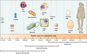
Cell size is limited by the surface area to volume ratio, which affects the cell's ability to transport materials efficiently. As cells grow larger, their volume increases faster than their surface area, limiting the rate of exchange with the environment.
Surface Area: Determines the amount of material that can enter or exit the cell.
Volume: Represents the space for cellular processes and metabolic activity.
Limiting Factor: If a cell becomes too large, the membrane cannot transport enough materials to support cellular functions.
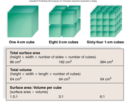
Formula: Surface area to volume ratio for a cube:
Types of Cells: Prokaryotic vs. Eukaryotic
Prokaryotic Cells
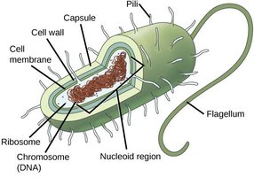
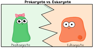
Prokaryotic cells are simpler and smaller than eukaryotic cells. They lack a membrane-bound nucleus and organelles.
Domains: Bacteria and Archaea
Features:
Circular DNA located in the nucleoid region
Cell wall, plasma membrane, ribosomes, cytoplasm
Eukaryotic Cells
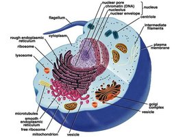
Eukaryotic cells are more complex, with a membrane-bound nucleus and various organelles. They belong to the domain Eukarya, which includes animals, plants, fungi, and protists.
Features:
Membrane-bound nucleus with chromosomes
Organelles such as mitochondria, endoplasmic reticulum, Golgi apparatus
Cytoplasm (cytosol)
Cellular Organelles and Their Functions
The Nucleus
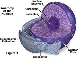
The nucleus is the control center of the cell, containing genetic information and coordinating cellular activities.
Nuclear Envelope: Double membrane surrounding the nucleus
Nuclear Pores: Regulate entry and exit of molecules
Nucleolus: Site of ribosome synthesis
Chromatin: DNA-protein complex forming chromosomes
Endoplasmic Reticulum (ER)
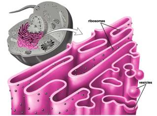
The ER is a network of membranes involved in protein and lipid synthesis. It is divided into rough and smooth types.
Rough ER: Studded with ribosomes; modifies proteins for secretion and sends transport vesicles to the Golgi apparatus
Smooth ER: Lacks ribosomes; synthesizes lipids and detoxifies drugs and poisons
Golgi Apparatus
The Golgi apparatus modifies, sorts, and packages proteins and lipids produced in the ER. It consists of flattened membrane sacs called cisternae.
Cis Face: Receives vesicles
Trans Face: Ships vesicles
Lysosomes and Peroxisomes
Lysosomes are membrane-bound sacs containing digestive enzymes, responsible for breaking down substances and cellular waste. Peroxisomes break down fatty acids and detoxify harmful substances.
Lysosomes: Digest cellular waste; involved in apoptosis (programmed cell death)
Peroxisomes: Oxidize fatty acids, producing hydrogen peroxide (); catalase enzyme breaks down $\text{H}_2\text{O}_2$
Ribosomes
Ribosomes are the site of protein synthesis, composed of rRNA and protein. They can be free in the cytoplasm or attached to the ER.
Function: Translate mRNA into amino acid chains (proteins)
Mitochondria
Mitochondria are the powerhouses of the cell, generating ATP through cellular respiration. They have a double membrane and their own DNA.
Matrix: Fluid-filled inner compartment
Endosymbiosis Theory: Suggests mitochondria originated from free-living prokaryotes
Chloroplasts
Chloroplasts are found in plant cells and are the site of photosynthesis. Like mitochondria, they have their own DNA and a double membrane.
Thylakoid Disks: Contain chlorophyll for capturing light energy
Endosymbiotic Theory
This theory explains the origin of mitochondria and chloroplasts in eukaryotic cells, proposing that they were once independent prokaryotes engulfed by ancestral eukaryotic cells.
Evidence:
Double-membrane structure
Own ribosomes and DNA
Reproduce independently within the cell
Vesicles and Vacuoles
Vesicles and vacuoles are membrane-bound structures used for storage and transport. Plant cells have a large central vacuole for storing water and nutrients.
Plasma Membrane
The plasma membrane is a phospholipid bilayer with embedded proteins, acting as a semipermeable barrier to regulate the movement of substances in and out of the cell.
Cell Wall
The cell wall is a rigid structure found in plant and bacterial cells, providing support and protection. In plants, it is composed of cellulose.
The Cytoskeleton and Cellular Movement
Cytoskeleton
The cytoskeleton is a network of fibers that provides structural support, maintains cell shape, and enables movement.
Microfilaments: Composed of actin; involved in movement
Microtubules: Hollow tubes; maintain shape and facilitate transport
Intermediate Filaments: Provide mechanical support
Cilia and Flagella
Cilia and flagella are cellular appendages used for movement. They have a "9+2" arrangement of microtubules.
Flagella: Long and few; propel cells through water
Cilia: Short and numerous; move fluids or enable locomotion
Centrosomes and Centrioles
Centrosomes are regions where microtubules originate, playing a key role in cell division. Centrioles are found in animal cells only.
Extracellular Structures and Cell Connections
Extracellular Matrix (ECM)
The ECM is found outside the plasma membrane of animal cells, composed of glycoproteins such as collagen. It strengthens tissues and transmits signals to cells.
Intercellular Junctions
Cells connect to each other through specialized junctions:
Plasmodesmata: Channels between plant cells
Tight Junctions: Seal cells together
Desmosomes: Anchor cells together
Gap Junctions: Allow communication between cells
Compartmentalization in Eukaryotic Cells
Importance of Internal Membranes
Eukaryotic cells have an extensive network of internal membranes that compartmentalize the cell, allowing different processes to occur in specialized environments. This organization contributes to the diversity and complexity of eukaryotic life.
Summary Table: Prokaryotic vs. Eukaryotic Cells
Feature | Prokaryotic Cells | Eukaryotic Cells |
|---|---|---|
Nucleus | No | Yes |
Organelles | No | Yes |
DNA | Circular, nucleoid region | Linear, in nucleus |
Domains | Bacteria, Archaea | Eukarya |
