 Indietro
Cell Trafficking and Cytoskeletal Structure: Study Notes for General Biology
Guida di studio - Note intelligenti
 Appunti personalizzati basati sui tuoi materiali, ampliati con definizioni chiave, esempi e contesto.
Cell Trafficking
Protein Targeting and Transport
Cells must direct proteins to specific locations to maintain proper function. This process, known as cell trafficking, involves a series of signals and pathways that ensure proteins reach their intended destinations, such as the cytoplasm, nucleus, mitochondria, chloroplast, endoplasmic reticulum (ER), Golgi apparatus, lysosome/vacuole, plasma membrane, or are secreted outside the cell.
Protein Traffic: Proteins are synthesized in the cytoplasm and may be directed to various organelles or exported from the cell.
Key Organelles: Cytoplasm, nucleus, mitochondria, chloroplast, ER, Golgi, lysosome/vacuole, plasma membrane.
Signal Sequences: Short amino acid sequences act as 'zip codes' to direct proteins to their correct location.
Nuclear Transport
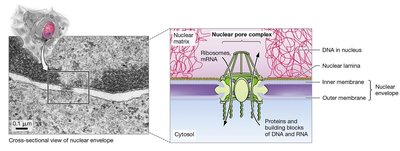
The nucleus is a site of constant molecular traffic, with proteins and nucleotides moving in and out through nuclear pores. Not all proteins of similar size can enter the nucleus; entry is determined by specific signals.
Nuclear Pore Complex: Large protein complexes that regulate traffic between the nucleus and cytoplasm.
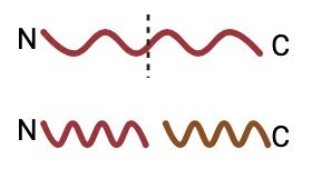
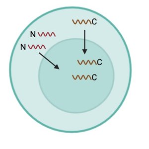
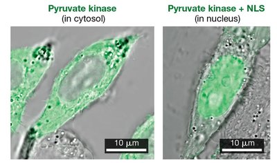
Nuclear Localization Signal (NLS): A ~17 amino acid sequence, often rich in lysine (K) and arginine (R), directs proteins into the nucleus.
Experiment (Nucleoplasmin): The tail region of nucleoplasmin contains the NLS, allowing nuclear entry, while the head does not.
Pulse-Chase Experiment: Tracking Protein Movement
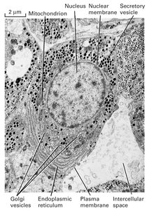
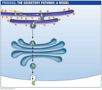
The pulse-chase experiment is used to track the movement of proteins through the secretory pathway. Proteins are labeled during a 'pulse' and their location is monitored over time ('chase').
Pulse: Cells are exposed to a labeled amino acid for a short time.
Chase: Cells are then exposed to unlabeled amino acids, and the movement of labeled proteins is tracked.
Results: Proteins move from the rough ER to the Golgi apparatus and then to secretory vesicles.
Protein Entry into the Rough ER
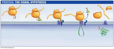
Proteins destined for secretion or for the endomembrane system are synthesized by ribosomes and enter the rough ER via a channel. The signal hypothesis explains how proteins are directed to the ER.
Signal Sequence: A short peptide at the N-terminus directs the ribosome to the ER membrane.
Translocation: The protein is threaded into the ER lumen as it is synthesized.
Protein Sorting and Vesicle Transport from the Golgi
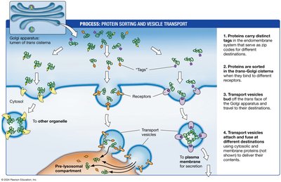
After proteins are processed in the Golgi apparatus, they are sorted and packaged into vesicles for transport to their final destinations.
Sorting Signals: Proteins carry distinct tags that determine their destination.
Vesicle Transport: Vesicles bud from the Golgi and fuse with target membranes.
Specificity: Receptors and tags ensure proteins are delivered accurately.
Cytoskeleton: Structure and Function
Overview of Cytoskeletal Elements
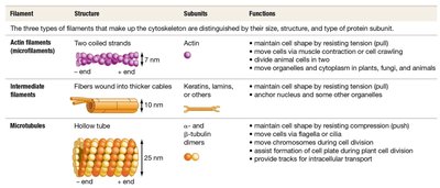
The cytoskeleton is a network of protein filaments that provides structure, shape, support, and facilitates movement within cells. It is composed of three main types of filaments: actin filaments, intermediate filaments, and microtubules.
Actin Filaments (Microfilaments): Responsible for cell shape, movement, and muscle contraction.
Intermediate Filaments: Provide structural support and anchor organelles.
Microtubules: Involved in intracellular transport, cell division, and movement of cilia/flagella.
Actin Filaments
Actin filaments are thin, flexible fibers involved in cell shape, movement, and muscle contraction. They can rapidly assemble and disassemble, allowing dynamic changes in cell structure.
Structure: Two coiled strands of actin, 7 nm in diameter.
Functions: Maintain cell shape, move cells via contraction or crawling, move organelles and cytoplasm.
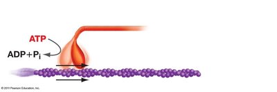
Movement: Motor proteins like myosin walk along actin filaments, using ATP for energy.
Intermediate Filaments
Intermediate filaments are thicker fibers made of various proteins (e.g., keratins, lamins) that provide structural support and anchor organelles.
Structure: Fibers wound into thicker cables, 10 nm in diameter.
Functions: Maintain cell shape by resisting tension, anchor nucleus and other organelles.
Examples: Keratins in cytoplasm, lamins in nucleus.
Microtubules
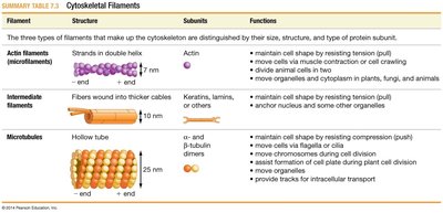
Microtubules are hollow tubes composed of α- and β-tubulin dimers. They are essential for intracellular transport, cell division, and movement of cilia and flagella.
Structure: Hollow tubes, 25 nm in diameter.
Functions: Maintain cell shape, move organelles, chromosomes, and provide tracks for motor proteins.
Motor Proteins: Kinesin and dynein move along microtubules, carrying cargo.
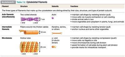
Summary Table: Cytoskeletal Filaments
The following table summarizes the structure, subunits, and functions of the three main cytoskeletal filaments:
Filament | Structure | Subunits | Functions |
|---|---|---|---|
Actin Filaments | Two coiled strands, 7 nm | Actin | Maintain cell shape, move cells, divide animal cells, move organelles |
Intermediate Filaments | Fibers wound into thicker cables, 10 nm | Keratins, lamins, others | Maintain cell shape, anchor nucleus and organelles |
Microtubules | Hollow tube, 25 nm | α- and β-tubulin dimers | Maintain cell shape, move cells, move chromosomes, assist cell division, provide tracks for transport |
Additional info:
Cell trafficking and cytoskeletal structure are fundamental to cell biology and are directly relevant to General Biology topics such as 'Inside the Cell', 'Protein Structure and Function', and 'Energy and Enzymes'.
Pulse-chase experiments and signal sequences are classic methods for studying protein movement and localization.
Cytoskeletal elements are critical for cell division, intracellular transport, and maintaining cell integrity.
