 Indietro
Cell–Cell Interactions: Structure, Communication, and Signaling
Guida di studio - Note intelligenti
 Appunti personalizzati basati sui tuoi materiali, ampliati con definizioni chiave, esempi e contesto.
Cell–Cell Interactions
Overview of Cell–Cell Interactions
Cell–cell interactions are fundamental for multicellular organisms, enabling cells to communicate, cooperate, and maintain tissue integrity. These processes involve both direct physical connections and signaling mechanisms that allow cells to respond to their environment and to each other.
Unicellular organisms adapt to environmental changes independently.
Multicellular organisms rely on interdependent communities of cells for coordinated function.
Chapter Roadmap
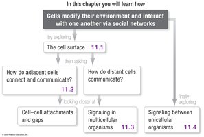
This chapter explores how cells modify their environment and interact via social networks, focusing on the cell surface, cell–cell attachments, and signaling mechanisms.
The Cell Surface
Structure of the Plasma Membrane
The plasma membrane is a phospholipid bilayer embedded with proteins, which regulate transport and facilitate cell–cell interactions.
Integral proteins: Span the membrane and are involved in transport and signaling.
Peripheral proteins: Attach to the membrane surface and cytoskeletal elements.
Membrane proteins create distinct internal environments and connect to extracellular structures.
The Extracellular Matrix (ECM) in Animals
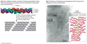
Most animal cells secrete an extracellular matrix (ECM), a fiber composite providing structural support and facilitating cell–cell communication.
Fibrous component: Mainly collagen, which forms triple helix fibrils for strength.
Ground substance: Composed of proteoglycans, giving cartilage its rubber-like consistency.
ECM composition and amount vary by tissue type; elastin allows tissues like lungs to stretch.
Integrins and ECM Attachment
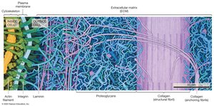
Integrins are membrane proteins that bind to ECM cross-linking proteins (e.g., laminins), anchoring the cytoskeleton to the ECM and connecting the plasma membrane to extracellular structures.
Cell–Cell Attachments and Communication
Overview of Cell–Cell Adhesion Structures
Direct physical connections between cells are essential for tissue structure and function. These attachments are especially important in epithelial tissues.
Tight Junctions
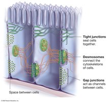
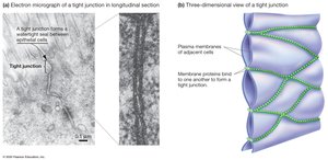
Tight junctions are cell–cell attachments composed of membrane proteins that form watertight seals between adjacent animal cells, primarily in epithelia. They are dynamic and can open or close in response to environmental changes.
Chains of proteins line up and bind, stitching cells together.
Regulate transport and maintain tissue integrity.
Desmosomes
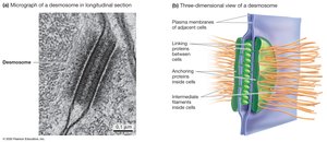
Desmosomes are strong cell–cell attachments common in epithelial and muscle cells. They consist of linking proteins and cytosolic anchoring proteins, reinforced by cytoskeletal intermediate filaments.
Provide mechanical strength to tissues.
Cadherins are the main linking proteins, binding only to cadherins of the same type.
Gap Junctions
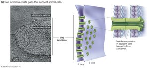
Gap junctions connect adjacent animal cells by forming protein channels, allowing the flow of ions and small molecules. These communication portals help coordinate activities and enable rapid passage of regulatory signals.
Directly connect cytoplasm of neighboring cells.
Regulate gene expression and protein activity.
Cell–Cell Signaling in Multicellular Organisms
Communication between Distant Cells
Cells not in physical contact communicate via signaling molecules, such as neurotransmitters and hormones. These molecules are secreted, circulate in the body, and act on target cells far from the signaling cell.
Neurotransmitters: Open or close channels in distant cells.
Hormones: Information-carrying molecules affecting target cells.
Signal Reception
Signal molecules deliver messages by binding to receptor proteins, which change shape and activity upon binding. Only cells with appropriate receptors respond to specific signaling molecules.
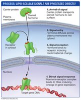
Lipid-soluble signals: Diffuse across plasma membrane; receptors are in cytoplasm.
Lipid-insoluble signals: Cannot cross membrane; receptors are on cell surface.
Receptors are dynamic and can be blocked (e.g., beta-blocker drugs).
Processing Lipid-Soluble Signaling Molecules
Lipid-soluble steroid hormones (e.g., estrogen, cortisol) diffuse through the plasma membrane, bind to cytoplasmic receptors, and form hormone–receptor complexes that enter the nucleus to alter gene expression.
Directly trigger changes in gene expression.
Cells produce different proteins in response.
Processing Lipid-Insoluble Signaling Molecules
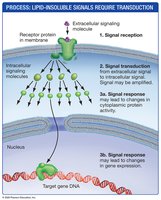
Lipid-insoluble signaling molecules cannot diffuse across the plasma membrane. They bind to cell surface receptors, triggering signal transduction—a process that converts extracellular signals to intracellular signals.
Signal transduction amplifies and diversifies the message.
Leads to changes in gene expression or protein activity.
Signal Transduction Pathways
G-Protein-Coupled Receptors
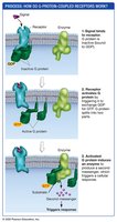
G-protein-coupled receptors (GPCRs) initiate production of intracellular second messengers, amplifying and diversifying the signal. G proteins are regulated by guanine nucleotides (GTP/GDP).
Signaling molecule binds to membrane receptor.
G protein exchanges GDP for GTP, activating the protein.
Active G protein subunit activates nearby enzyme, producing a second messenger.
Second messengers diffuse rapidly and amplify the signal.
Protein kinases may be activated, adding phosphate groups to proteins.
Enzyme-Linked Receptors
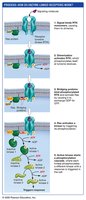
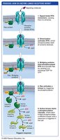
Enzyme-linked receptors, such as receptor tyrosine kinases (RTKs), directly catalyze reactions inside the cell. RTK signal transduction involves dimerization, autophosphorylation, and activation of a phosphorylation cascade.
Hormone binds to two RTK subunits, forming a dimer.
RTK phosphorylates itself using ATP.
Proteins bridge RTK and Ras protein, activating Ras.
Activated Ras triggers a phosphorylation cascade of protein kinases.
Mitogen-activated protein kinases (MAPKs) are key in cell division signaling.
Signal Deactivation
Cells possess mechanisms for rapid signal deactivation, such as phosphatases removing phosphate groups from proteins. This ensures sensitivity to small changes in signaling.
Crosstalk: Integration of Multiple Signals
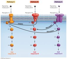
Signal transduction pathways intersect and connect, forming complex networks that allow cells to respond in an integrated manner. Crosstalk can inhibit or stimulate steps in other pathways, providing multiple points for regulation.
One pathway may inhibit another, reducing response.
One pathway may stimulate another, leading to diverse responses.
Multiple steps allow for extensive crosstalk.
Summary Table: Cell–Cell Attachment Structures
Structure | Main Function | Key Components | Example Location |
|---|---|---|---|
Tight Junction | Watertight seal between cells | Membrane proteins | Epithelial tissue |
Desmosome | Strong adhesion, mechanical strength | Cadherins, intermediate filaments | Epithelium, muscle |
Gap Junction | Communication portal, molecule exchange | Channel proteins | Animal tissues |
Key Equations and Concepts
Phosphorylation Reaction:
GTP Hydrolysis:
Additional info: Signal transduction pathways are highly conserved across eukaryotes, and defects in these pathways are linked to diseases such as cancer and diabetes.
