 Indietro
Cellular Basis of Reproduction and Inheritance: Study Notes
Guida di studio - Note intelligenti
 Appunti personalizzati basati sui tuoi materiali, ampliati con definizioni chiave, esempi e contesto.
Cell Division and Reproduction
Introduction to Cellular Division
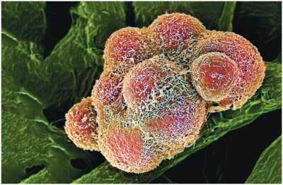
Cell division is a fundamental process required for growth, maintenance, and reproduction in all living organisms. While uncontrolled cell division leads to cancer, normal cell division is essential for life.
Cancer cells: Undergo uncontrolled cell division, disrupting normal tissue function.
Normal cell division: Necessary for organismal development, tissue repair, and reproduction.
Types of Reproduction
Organisms reproduce either asexually or sexually, each method having distinct genetic consequences.
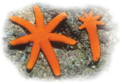
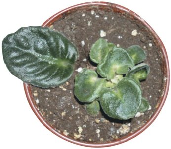
Asexual reproduction: Produces offspring genetically identical to the parent (clones).
Examples: Fragmentation in sea stars, plant cuttings.
Sexual reproduction: Produces offspring with unique gene combinations via fusion of gametes.
Gametes: Produced by reductive cell division (meiosis).
Importance of Cell Division
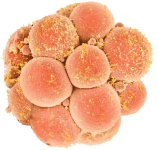
Cell division is vital for multicellular organisms, enabling growth from a single cell and replacement of damaged or lost cells.
Growth: From a single zygote to trillions of cells.
Repair: Millions of cells divide every second to replace damaged or lost cells.
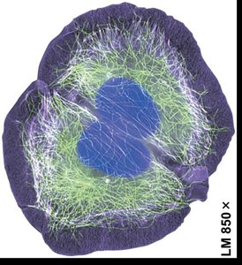
Cell Division in Prokaryotic Cells
Binary Fission
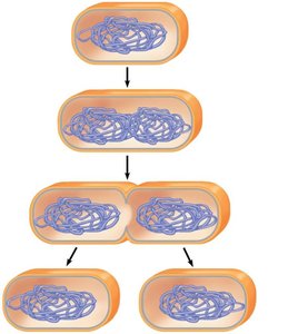
Prokaryotes reproduce by binary fission, a process that divides a cell in half to produce two daughter cells.
Binary fission: Simple, efficient method of reproduction in prokaryotes.
Cell wall: Prokaryotes possess cell walls (not made of cellulose).
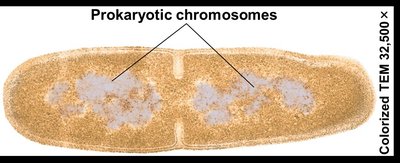
Steps of Binary Fission
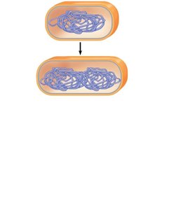
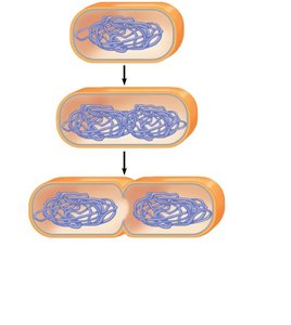
Binary fission involves chromosome duplication, cell elongation, and division into two daughter cells.
Step 1: Chromosome duplication and separation.
Step 2: Continued elongation and movement of chromosome copies.
Step 3: Division into two daughter cells.
The Eukaryotic Cell Cycle and Mitosis
Chromosomes and Chromatin
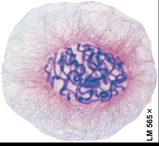
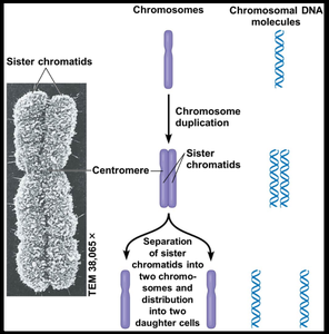
Eukaryotic cells contain multiple chromosomes housed within the nucleus. Chromosomes are composed of DNA and proteins, forming chromatin.
Chromatin: Loosely packed DNA-protein complex, visible only when condensed.
Chromosome duplication: Produces sister chromatids with identical DNA.
The Cell Cycle
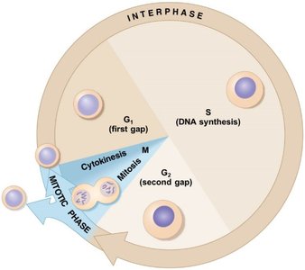
The cell cycle is an ordered sequence of events from cell formation to its own division. It includes interphase (G1, S, G2) and mitotic phase (mitosis and cytokinesis).
Interphase: Cell grows (G1), duplicates DNA (S), prepares for division (G2).
Mitotic phase: Mitosis (nuclear division) and cytokinesis (cytoplasmic division).
Stages of Mitosis
Mitosis is divided into five main stages: prophase, prometaphase, metaphase, anaphase, and telophase. Chromosome movement is driven by the mitotic spindle.
Prophase: Chromosomes condense, spindle forms.
Prometaphase: Nuclear envelope dissolves, spindle attaches to kinetochores.
Metaphase: Chromosomes align at the metaphase plate.
Anaphase: Sister chromatids separate and move to opposite poles.
Telophase: Nuclear envelope reforms, chromosomes decondense.
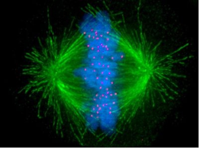
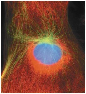
Interphase and Prophase
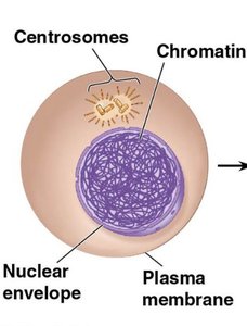
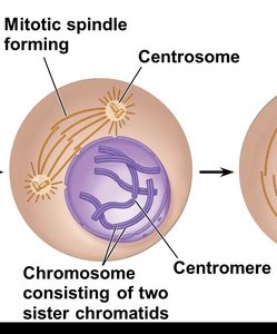
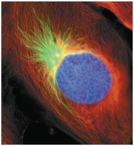
During interphase, chromosomes duplicate and nucleoli are visible. In prophase, chromosomes condense and the mitotic spindle begins to form.
Interphase: Chromosomes in chromatin form, nuclear envelope intact.
Prophase: Chromosomes visible, spindle forms.
Control of Cell Division
Regulation Mechanisms
Cell division is tightly regulated to ensure proper growth, development, and maintenance. Cells require anchorage and density-dependent inhibition to divide.
Anchorage dependency: Cells must contact a solid surface to divide.
Density-dependent inhibition: Crowding stops cell division.
Cancer cells: Ignore these controls, divide uncontrollably.
Growth Factors and Checkpoints
Chemical signals called growth factors trigger cell division. The cell cycle is regulated by checkpoints influenced by these factors.
Growth factors: Bind to receptors to initiate division.
G0 phase: Non-dividing state (e.g., mature nerve and muscle cells).
Meiosis: Reduction Division
Chromosome Number and Homologous Pairs
Somatic cells contain a specific number of chromosomes arranged in homologous pairs. Gametes are haploid, containing a single set of chromosomes.
Diploid (2n): Two sets of chromosomes (somatic cells).
Haploid (n): One set of chromosomes (gametes).
Homologous chromosomes: Carry genes for the same traits at the same loci.
Meiosis Process
Meiosis involves two consecutive divisions, resulting in four haploid cells. Meiosis I separates homologous pairs; Meiosis II separates sister chromatids.
Meiosis I: Homologous chromosomes separate.
Meiosis II: Sister chromatids separate.
Crossing over: Exchange of genetic material between homologous chromosomes during Prophase I.
Comparison of Mitosis and Meiosis
Mitosis produces two genetically identical diploid cells, while meiosis produces four genetically unique haploid cells.
Mitosis: One division, no crossing over, identical daughter cells.
Meiosis: Two divisions, crossing over, unique daughter cells.
Process | Number of Divisions | Genetic Variation | Resulting Cells |
|---|---|---|---|
Mitosis | 1 | No | 2 diploid, identical |
Meiosis | 2 | Yes (crossing over, independent assortment) | 4 haploid, unique |
Sources of Genetic Variation
Independent Assortment
During meiosis, chromosomes are randomly distributed to gametes, resulting in numerous possible combinations.
Formula: where n = number of homologous pairs.
Humans: possible combinations per gamete.
Random Fertilization
The fusion of gametes from two parents further increases genetic diversity, producing over 70 trillion possible zygote combinations in humans.
Calculation: possible zygote combinations.
Crossing Over
Crossing over during Prophase I of meiosis exchanges segments between nonsister chromatids, creating new genetic combinations.
Chiasma: Site of crossing over.
Result: Recombinant chromosomes with new gene combinations.
Alterations of Chromosome Number and Structure
Nondisjunction
Nondisjunction is the failure of chromosomes to separate properly during meiosis, resulting in abnormal chromosome numbers.
Meiosis I: Homologous chromosomes fail to separate.
Meiosis II: Sister chromatids fail to separate.
Karyotype Analysis
A karyotype is a photographic inventory of chromosomes, used to detect abnormalities such as trisomy 21 (Down syndrome).
Trisomy 21: Most common chromosome number abnormality.
Maternal age: Increased risk of Down syndrome with maternal age.
Sex Chromosome Abnormalities
Nondisjunction of sex chromosomes can result in syndromes such as Klinefelter (XXY), Turner (XO), and others.
Sex Chromosomes | Syndrome | Frequency |
|---|---|---|
XXY | Klinefelter (male) | 1 / 2,000 |
XYY | Normal male | 1 / 2,000 |
XXX | Normal female | 1 / 1,000 |
XO | Turner (female) | 1 / 5,000 |
Polyploidy and Chromosome Structure Alterations
Polyploid organisms have extra sets of chromosomes, which can lead to new species. Structural alterations (deletions, duplications, translocations) can cause birth defects and cancer.
Example: Gray tree frog (Hyla versicolor) is tetraploid.
Translocation: Can activate cancer-causing genes (e.g., chronic myelogenous leukemia).
