 Indietro
Chapter 37: Neurons, Synapses, and Signaling – Study Notes
Guida di studio - Note intelligenti
 Appunti personalizzati basati sui tuoi materiali, ampliati con definizioni chiave, esempi e contesto.
Neuron, Synapses, and Signaling
Overview: Lines of Communication
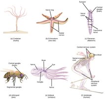
The nervous system is responsible for communication within the body, utilizing specialized cells called neurons. Neurons transmit information using two types of signals: electrical signals for long-distance communication and chemical signals for short-distance communication. The interpretation and processing of these signals occur in clusters of neurons called ganglia or in more complex structures such as the brain.
Concept 37.1: Neuron Structure and Organization
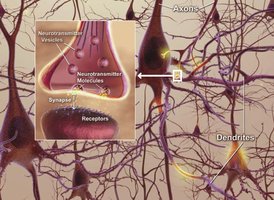
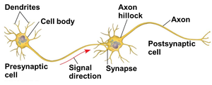
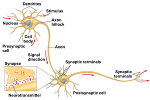
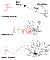
Neurons exemplify the close relationship between structure and function, a result of evolutionary adaptation. The main parts of a neuron include the cell body (containing most organelles), dendrites (branched extensions that receive signals), and a single axon (long extension that transmits signals). The axon hillock is the region where signals are generated, and the synapse is the junction where axons transmit signals to other cells, often using neurotransmitters.
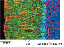
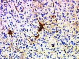
Supporting cells called glial cells are essential for neuron function. In the mammalian brain, glia outnumber neurons by 10- to 50-fold, providing structural and metabolic support.
Introduction to Information Processing
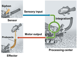
Nervous systems process information in three stages: sensory input, integration, and motor output. Sensory neurons transmit information about external or internal stimuli to processing centers. Interneurons integrate and interpret this input, while motor neurons transmit signals to effectors (muscles or glands) to elicit a response.
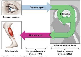
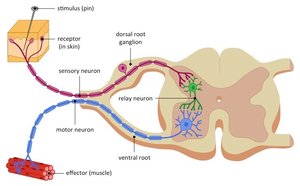
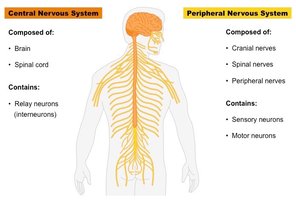
The central nervous system (CNS) consists of neurons that carry out integration, while the peripheral nervous system (PNS) includes neurons that carry information into and out of the CNS. Bundles of PNS neurons form nerves.
Concept 37.2: Ion Pumps and Ion Channels Establish the Resting Potential
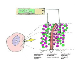
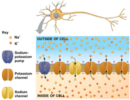
The inside of a neuron is negatively charged relative to the outside, creating a membrane potential. The resting potential is the membrane potential of a neuron not sending signals. Changes in membrane potential serve as signals for transmitting and processing information.
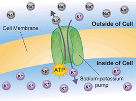
Potassium (K+) and Sodium (Na+) ions play key roles in establishing the resting potential. The sodium-potassium pump uses ATP to maintain K+ and Na+ gradients across the plasma membrane.
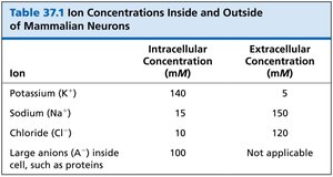
Ion | Intracellular Concentration (mM) | Extracellular Concentration (mM) |
|---|---|---|
Potassium (K+) | 140 | 5 |
Sodium (Na+) | 15 | 150 |
Chloride (Cl-) | 10 | 120 |
Large anions (A-) inside cell, such as proteins | 100 | Not applicable |
Ion channels are selectively permeable, allowing only certain ions to pass. A resting neuron has many open potassium channels, allowing K+ to flow out, which contributes to the negative charge inside.
Modeling the Resting Potential
The resting potential can be modeled by an artificial membrane separating two chambers with different KCl concentrations. K+ diffuses down its gradient, and negative charge builds up in the inner chamber. At equilibrium, both electrical and chemical gradients are balanced. The equilibrium potential (Eion) for an ion can be calculated using the Nernst equation:
For K+, EK ≈ –90 mV; the actual resting potential is about –60 to –80 mV due to some Na+ influx.
Concept 37.3: Action Potentials
Neurons contain gated ion channels that open or close in response to stimuli. Voltage-gated ion channels respond to changes in membrane voltage. When a depolarization reaches threshold, an action potential is generated—a rapid, all-or-none change in membrane voltage that transmits signals over long distances.
Hyperpolarization and Depolarization
Opening K+ channels causes hyperpolarization (membrane potential becomes more negative). Opening Na+ channels causes depolarization (membrane potential becomes less negative).
Graded Potentials and Action Potentials
Graded potentials are changes in polarization that vary with stimulus strength and decay with distance. If depolarization is sufficient, an action potential occurs.
Generation of Action Potentials: Stages
Resting state: Most voltage-gated Na+ and K+ channels are closed.
Depolarization: Some Na+ channels open, Na+ enters.
Rising phase: Threshold crossed, membrane potential increases rapidly.
Falling phase: Na+ channels inactivate, K+ channels open, K+ exits.
Undershoot: K+ permeability is high, then returns to rest.
During the refractory period, a second action potential cannot be initiated due to Na+ channel inactivation.
Conduction of Action Potentials
Action potentials are initiated at the axon hillock and travel toward synaptic terminals. Inactivated Na+ channels prevent backward propagation.
Evolutionary Adaptations of Axon Structure
Action potential speed increases with axon diameter. In vertebrates, myelin sheaths (produced by glia) insulate axons, enabling fast conduction via saltatory conduction (action potentials jump between nodes of Ranvier).
Concept 37.4: Neurons Communicate at Synapses
Most synapses are chemical synapses, where neurotransmitters carry information from the presynaptic neuron to the postsynaptic cell. Neurotransmitters are packaged in synaptic vesicles and released upon arrival of an action potential, diffusing across the synaptic cleft to bind receptors on the postsynaptic cell.
Generation of Postsynaptic Potentials
Neurotransmitter binding to ligand-gated ion channels generates postsynaptic potentials:
Excitatory postsynaptic potentials (EPSPs): Depolarizations that bring membrane potential toward threshold.
Inhibitory postsynaptic potentials (IPSPs): Hyperpolarizations that move membrane potential farther from threshold.
Summation of Postsynaptic Potentials
Postsynaptic potentials can combine via temporal summation (rapid succession) or spatial summation (simultaneous input from multiple synapses). The combined effect determines whether an action potential is triggered.
Modulated Signaling at Synapses
Some neurotransmitters bind to metabotropic receptors, activating signal transduction pathways and second messengers. These effects are slower but longer-lasting than ligand-gated channels.
Acetylcholine
Acetylcholine is a common neurotransmitter involved in muscle stimulation, memory, and learning. It acts via ligand-gated and metabotropic receptors. Toxins such as sarin and botulinum disrupt acetylcholine signaling.
Example: At the vertebrate neuromuscular junction, acetylcholine released by motor neurons binds to ligand-gated ion channels, generating an EPSP and triggering muscle contraction.
Additional info: There are over 100 known neurotransmitters, each with specific functions and receptor types.
----------------------------------------
