 Indietro
Circulation and Gas Exchange: Structure and Function of Circulatory and Respiratory Systems
Guida di studio - Note intelligenti
 Appunti personalizzati basati sui tuoi materiali, ampliati con definizioni chiave, esempi e contesto.
Circulatory and Respiratory Systems Overview
Introduction to Circulatory and Respiratory Systems
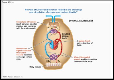
The circulatory and respiratory systems are essential for the transport and exchange of gases, nutrients, and wastes in multicellular organisms. The circulatory system consists of the heart and blood vessels, while the respiratory system includes the lungs and airways. These systems work together to maintain homeostasis and support cellular metabolism.
Circulatory System: Heart and blood vessels (arteries, veins, capillaries).
Respiratory System: Lungs and airways (trachea, bronchi, bronchioles).
Function: Exchange and transport of oxygen (O2), carbon dioxide (CO2), nutrients, and wastes.
Surface Area to Volume Ratio
Importance of Surface Area to Volume Ratio
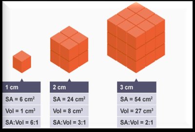
The surface area to volume ratio is a critical factor in biological systems, influencing the efficiency of material exchange. As organisms increase in size, their surface area relative to volume decreases, affecting diffusion rates.
Surface Area: Total area exposed to the environment.
Volume: Internal space of the organism.
Key Principle: Smaller objects have a higher surface area to volume ratio, facilitating faster diffusion.
Factors Increasing SA:Vol Ratio: Small size, thin/flat shape, folding, branching, elongated structures.
Diffusion Rate and Exchange in Small Organisms
Small unicellular and simple multicellular organisms rely on direct diffusion for gas and nutrient exchange due to their high surface area to volume ratio.
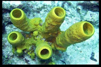
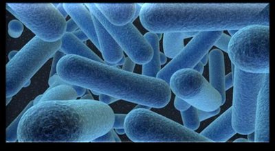
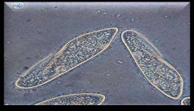
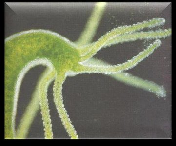
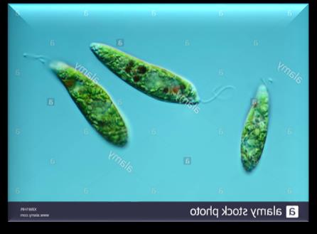
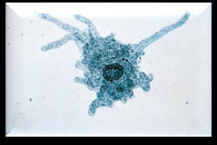
Direct Diffusion: Efficient in bacteria, protists, protozoa, hydra, and sponges.
Examples:
Bacteria
Sponges
Paramecium
Hydra
Euglena
Amoeba
Types of Circulatory Systems
Open vs Closed Circulatory Systems
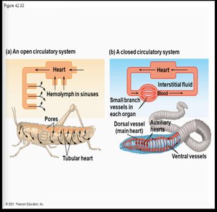
Larger multicellular organisms require specialized vasculature for efficient transport. There are two main types of circulatory systems: open and closed.
Open Circulatory System:
No capillaries; hemolymph bathes organs directly.
Found in arthropods (insects, crustaceans) and mollusks (clams, snails).
Contains hemocyanin (copper-based oxygen carrier).
Closed Circulatory System:
Capillaries present; blood confined to vessels.
Found in vertebrates (mammals, birds, reptiles, amphibians, fish).
Vascular Tissues in Mammals
Structure and Function of Blood Vessels
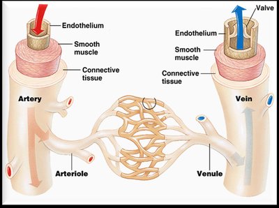
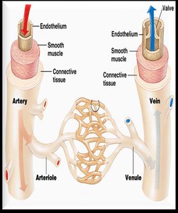
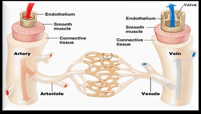
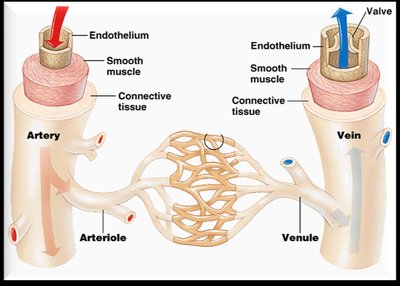
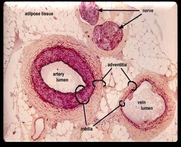
Mammalian blood vessels are classified as arteries, arterioles, veins, venules, and capillaries, each with distinct histological features and functions.
Arteries: Thick-walled, elastic, carry blood away from the heart; high blood pressure.
Arterioles: Smaller branches of arteries; regulate blood flow within organs.
Veins: Thin-walled, larger diameter, carry blood to the heart; low blood pressure; contain valves.
Venules: Small veins merging from capillaries.
Capillaries: Microscopic vessels for exchange; composed of endothelial cells and basal lamina.
Capillaries and Exchange of Materials
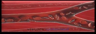
Capillaries are the primary site for the exchange of gases, nutrients, and wastes between blood and tissues.
Structure: Single layer of endothelial cells; diameter allows single-file passage of erythrocytes.
Function: Exchange of O2, CO2, amino acids, monosaccharides, and nitrogenous wastes.
Interstitial Fluid (ISF): Fluid surrounding tissue cells; mediates exchange between capillaries and cells.
The Mammalian Heart
Heart Structure and Blood Flow
The mammalian heart is a four-chambered organ responsible for pumping blood through two circuits: pulmonary and systemic.
Chambers: Two atria (receive blood), two ventricles (pump blood).
Valves: Ensure unidirectional flow; include atrioventricular (tricuspid, bicuspid) and semilunar valves.
Blood Vessels:
Vena cavae: bring deoxygenated blood to right atrium.
Pulmonary artery: carries deoxygenated blood to lungs.
Pulmonary veins: return oxygenated blood to left atrium.
Aorta: distributes oxygenated blood to body.
Blood Flow in Circulatory Circuits
Systemic and Pulmonary Circuits
Blood circulates through two main circuits:
Systemic Circuit: Heart → body tissues → heart
Pulmonary Circuit: Heart → lungs → heart
Direction of Blood Flow in Systemic Circuit: aorta → arteries → arterioles → capillaries → venules → veins → vena cava Direction of Blood Flow in Pulmonary Circuit: pulmonary artery → pulmonary arterioles → pulmonary capillaries → pulmonary venules → pulmonary veins
The Cardiac Cycle
Phases of the Cardiac Cycle
The cardiac cycle describes the sequence of heart contraction and relaxation.
Sinoatrial Node (SA): Pacemaker; initiates electrical impulses.
Phases:
Diastole: Heart relaxed; blood flows into atria and ventricles.
Atrial systole: Atria contract; blood forced into ventricles.
Ventricular systole: Ventricles contract; blood pumped out.
Electrocardiogram (ECG): Measures electrical activity; P wave (atrial systole), QRS wave (ventricular systole), T wave (ventricular diastole).
Blood Pressure
Measurement and Regulation of Blood Pressure
Blood pressure is the force exerted by blood on vessel walls, measured in mmHg.
Systolic Pressure: Pressure during ventricular contraction.
Diastolic Pressure: Pressure during ventricular relaxation.
Normal BP: 120/70–80 mmHg
Hypertension: ≥140/90 mmHg
Sphygmomanometer: Instrument for measuring blood pressure.
Heart Attacks and Strokes
Atherosclerosis, Myocardial Infarction, and Stroke
Atherosclerosis: Hardening of arteries due to fatty deposits; can lead to blockage.
Myocardial Infarction: Heart attack; blocked coronary arteries cause heart cell death.
Stroke: Blockage in arteries of neck/head; causes death of nervous tissue.
Blood Pressure and Velocity in Capillaries
Pressure and Velocity Changes
Blood Pressure: Highest in arteries, decreases in arterioles and capillaries.
Blood Velocity: Slowest in capillaries, increases in veins.
Boyle’s Law: (Pressure is inversely proportional to volume in a closed system).
Sphincters and Valves
Control of Blood Flow
Sphincter: Ring of smooth muscle; controls passage between locations.
Valve: Ensures unidirectional flow of blood.
Precapillary Sphincters: Regulate blood distribution in capillary beds.
Blood Composition
Types of Blood Cells and Plasma
Blood consists of cells and plasma, each with specific functions.
Erythrocytes: Red blood cells; contain hemoglobin.
Leukocytes: White blood cells; five types (neutrophils, eosinophils, basophils, monocytes, lymphocytes).
Plasma: Extracellular matrix; ~90% water, 10% nutrients, wastes, hormones, ions, proteins.
Functions: Transport, regulation, protection.
Respiratory System
Respiratory Surfaces and Gas Exchange Mechanisms
Gas exchange occurs across specialized surfaces, with mechanisms varying by organism.
Direct Diffusion: Small organisms; high surface area.
Highly Vasculated Skin: Earthworms, amphibians.
Tracheae: Arthropods; air tubes for gas exchange.
Gills: Aquatic animals; countercurrent exchange.
Lungs: Higher vertebrates; alveoli provide large surface area.
Alveoli and Surfactant
Alveoli: Microscopic air sacs; one cell thick for efficient diffusion.
Pneumocytes: Cell type in alveoli; type II secrete surfactant.
Surfactant: Reduces surface tension; prevents alveolar collapse.
Respiratory Distress Syndrome (RDS): Alveoli collapse; common in premature infants.
Mechanics and Control of Breathing
Muscles and Negative Air Pressure
Intercostal Muscles: Between ribs; aid in breathing.
Diaphragm: Skeletal muscle; separates thoracic and abdominal cavities.
Boyle’s Law: (Pressure decreases as volume increases).
Inhalation: Active; muscles contract, thoracic volume increases, pressure decreases.
Exhalation: Passive; muscles relax, thoracic volume decreases, pressure increases.
Control of Breathing Rate
Medulla and Pons: Brainstem regions; regulate breathing.
Chemoreceptors: Sense CO2, H+, and pH in blood and cerebrospinal fluid.
Equation:
Gases and Partial Pressure
Partial Pressure and Gas Exchange
Partial pressure is the pressure exerted by a specific gas in a mixture. Gases diffuse from areas of higher to lower partial pressure.
PPO2 (atmosphere): 160 mmHg
PPO2 (alveoli): 104 mmHg
PPO2 (capillaries): 40 mmHg
PPCO2 (atmosphere): 0.03 mmHg
PPCO2 (alveoli): 40 mmHg
PPCO2 (capillaries): 45 mmHg
Hemoglobin and Oxygen Transport
Structure and Function of Hemoglobin
Hemoglobin is a quaternary protein in red blood cells responsible for oxygen transport.
Structure: Four polypeptide chains (2 alpha, 2 beta); each with a heme group and iron.
Function: Each hemoglobin binds up to four O2 molecules.
Myoglobin: Muscle protein; binds one O2 molecule.
Carbon Dioxide Transport
CO2 Transport and Buffering
At Tissues: CO2 enters RBCs, forms H2CO3 (carbonic acid), dissociates to H+ and HCO3-.
Hemoglobin: Buffers blood pH by binding H+.
At Lungs: HCO3- re-enters RBCs, forms CO2 for exhalation.
Loading and Unloading of Oxygen and Bohr Shift
Oxygen Saturation and Bohr Effect
Hemoglobin loads and unloads O2 based on partial pressure and pH.
At Lungs: High PPO2; hemoglobin is 98% saturated.
At Tissues: Lower PPO2; hemoglobin unloads O2.
Bohr Shift: Increased CO2 lowers pH, decreases hemoglobin affinity for O2, promoting O2 release.
Summary Table: Circulatory System Components
Component | Structure | Function |
|---|---|---|
Artery | Thick, elastic walls | Carry blood away from heart |
Vein | Thin walls, valves | Carry blood to heart |
Capillary | Single layer of endothelium | Exchange of gases, nutrients, wastes |
Heart | Four chambers, valves | Pump blood through circuits |
Summary Table: Respiratory System Mechanisms
Mechanism | Organism Type | Structure |
|---|---|---|
Direct Diffusion | Unicellular, simple multicellular | Cell membrane |
Vasculated Skin | Earthworm, amphibians | Moist skin |
Tracheae | Arthropods | Air tubes |
Gills | Aquatic animals | Gill filaments, lamellae |
Lungs | Mammals, birds, reptiles | Alveoli |
