 Indietro
BIO 102 Chapter 42 Part 1 A
Guida di studio - Note intelligenti
 Appunti personalizzati basati sui tuoi materiali, ampliati con definizioni chiave, esempi e contesto.
BIO 102 Chapter 42 Part 1
Physical Constraints and Exchange in Multicellular Organisms
Multicellular organisms face physical constraints that limit the efficiency of diffusion for transporting substances across large distances. To overcome these limitations, they pair specialized high surface area exchange surfaces with bulk flow fluid transport systems. This integrative approach connects foundational concepts such as surface area-to-volume relationships, cell signaling, and endocrine regulation to the function of circulatory and respiratory systems.
Exchange of Materials in Organisms
Trading Places: The Role of Circulatory System in Material Exchange
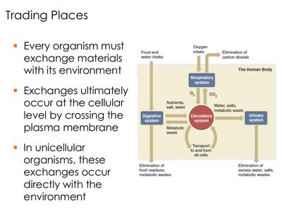
Every organism must exchange materials with its environment, a process that occurs at the cellular level by crossing the plasma membrane. In unicellular organisms, exchanges occur directly with the environment, while multicellular organisms rely on specialized systems for transport and elimination of metabolic waste.
Key Point 1: Exchange of substances is essential for survival and occurs across plasma membranes.
Key Point 2: The circulatory system integrates with other systems (urinary, digestive, respiratory) to transport and eliminate waste.
Example: The circulatory system delivers metabolic waste to the urinary system for elimination.
Specialized Exchange Surfaces: Gills in Amphibians
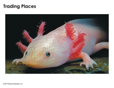
Some organisms, such as amphibians, utilize specialized structures like gills for gas exchange. Gills increase surface area, enhancing the efficiency of oxygen uptake and carbon dioxide elimination.
Key Point 1: Gills provide a high surface area for exchange of O2 and CO2.
Key Point 2: Oxygen serves as the final electron acceptor in cellular respiration, supporting ATP production.
Example: The axolotl uses feathery gills to maximize gas exchange with its aquatic environment.
Diffusion and Circulatory Systems
Concept 42.1: Circulatory Systems Link Exchange Surfaces with Cells
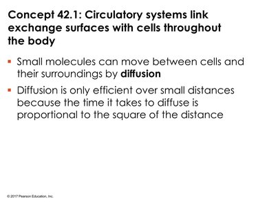
Small molecules move between cells and their surroundings by diffusion, which is efficient only over microscopic distances. The time required for diffusion increases with the square of the distance, making it insufficient for large organisms.
Key Point 1: Diffusion is a passive process driven by concentration gradients.
Key Point 2: Efficiency of diffusion decreases rapidly as distance increases.
Formula: (Time to diffuse is proportional to the square of the distance)
Gastrovascular Cavities in Simple Animals
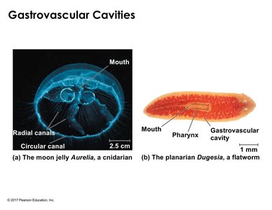
Some simple animals lack circulatory systems and instead use gastrovascular cavities for both digestion and distribution of substances. These cavities are typically thin, facilitating efficient diffusion.
Key Point 1: Gastrovascular cavities combine digestive and circulatory functions.
Key Point 2: Thin body walls support efficient diffusion across the cavity.
Example: Aurelia (moon jelly) and Dugesia (planarian flatworm) utilize gastrovascular cavities.
Types of Circulatory Systems
Open vs. Closed Circulatory Systems
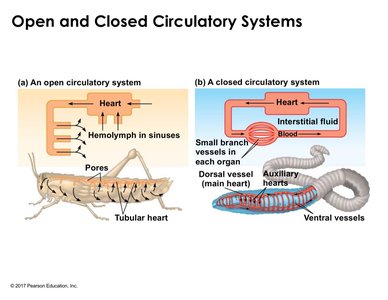
Circulatory systems consist of a circulatory fluid, vessels, and a muscular pump (heart). Open systems, found in arthropods, use hemolymph that bathes organs directly, while closed systems, found in annelids and vertebrates, keep blood enclosed within vessels.
Key Point 1: Open systems lack distinction between circulatory and interstitial fluid; closed systems maintain separation.
Key Point 2: Closed systems allow for more efficient and directed transport of substances.
Comparison Table:
System Type | Circulatory Fluid | Vessel Structure | Example Organism |
|---|---|---|---|
Open | Hemolymph | Sinuses, open vessels | Grasshopper |
Closed | Blood | Enclosed vessels | Earthworm |
Vertebrate Circulatory System Organization
Cardiovascular System Structure
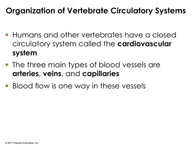
Humans and other vertebrates possess a closed circulatory system known as the cardiovascular system. It consists of three main types of blood vessels: arteries, veins, and capillaries. Blood flow is unidirectional, ensuring efficient transport.
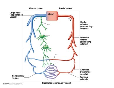
Key Point 1: Arteries carry blood away from the heart (efferent), veins return blood to the heart (afferent).
Key Point 2: Capillaries serve as exchange vessels between arteries and veins.
Example: The aorta is a major elastic artery, while the femoral artery is a muscular artery.
Blood Vessel Types and Functions
Blood vessels are classified based on their structure and function. Elastic arteries conduct blood under high pressure, muscular arteries distribute blood to specific regions, arterioles regulate flow to tissues, and capillaries facilitate exchange. Veins serve as reservoirs and return blood to the heart.
Key Point 1: Elastic arteries withstand high pressure; muscular arteries regulate flow via vasoconstriction.
Key Point 2: Veins have larger lumens and thinner walls compared to arteries.
Example: Capillary beds are sites of nutrient and gas exchange.
Circulatory Patterns in Vertebrates
Single Circulation in Fish
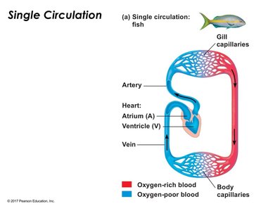
Fish possess a single circulatory loop, with a two-chambered heart (one atrium, one ventricle). Blood flows from the heart to gill capillaries for oxygenation, then to body capillaries, and returns to the heart. This system limits pressure and oxygen delivery to tissues.
Key Point 1: Single circulation results in lower blood pressure after passing through gills.
Key Point 2: Oxygen-poor blood returns to the heart for reoxygenation.
Example: Fish utilize single circulation for transport of oxygen and nutrients.
Double Circulation in Amphibians and Mammals
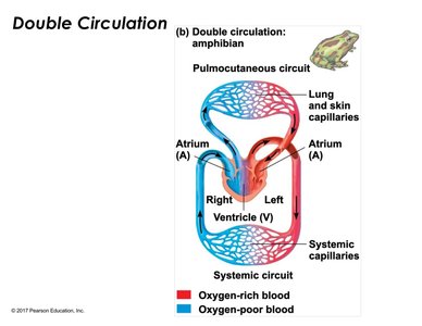
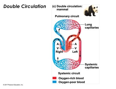
Double circulation separates pulmonary and systemic circuits, allowing for higher pressure and more efficient oxygen delivery. Amphibians have a three-chambered heart, while mammals have a four-chambered heart, completely separating oxygen-rich and oxygen-poor blood.
Key Point 1: Double circulation maintains higher blood pressure in organs.
Key Point 2: Mammals' four-chambered heart supports endothermy and high metabolic rates.
Example: Amphibians use both lungs and skin for oxygenation; mammals use lungs exclusively.
Heart Structure and Function
Concept 42.2: Coordinated Cycles of Heart Contraction
The mammalian cardiovascular system is organized to meet the body's continuous demand for oxygen, supporting ATP production through cellular respiration.
Key Point 1: Heart contraction cycles drive double circulation.
Key Point 2: Oxygen is essential for ATP generation in cells.
Anatomy of the Mammalian Heart
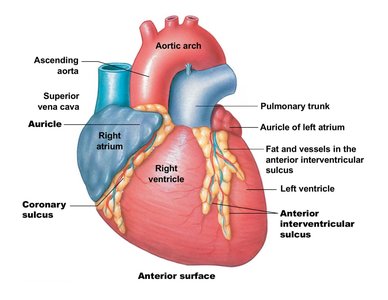
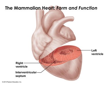
The heart is located in the thoracic cavity and consists of four chambers: right and left atria, right and left ventricles. The atria receive blood, while the ventricles distribute it. Surface features include the coronary sulcus and interventricular sulcus, which mark boundaries between chambers.
Key Point 1: The right ventricle is primarily responsible for pulmonary circulation; the left ventricle for systemic circulation.
Key Point 2: The left ventricle has a thicker wall to generate higher pressure for systemic circulation.
Example: The coronary sulcus separates atria from ventricles.
A Closer Look at Heart Chambers and Valves
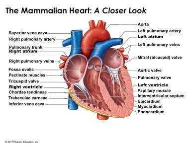
The heart contains atrioventricular (AV) valves (tricuspid and bicuspid/mitral) and semilunar valves (pulmonary and aortic). Valves prevent backflow and open/close in response to pressure changes. The heart wall consists of three layers: epicardium, myocardium, and endocardium.
Key Point 1: AV valves separate atria from ventricles; semilunar valves control exit from ventricles.
Key Point 2: The myocardium is responsible for heart contractions.
Example: The fossa ovalis is a remnant of fetal circulation found in the interatrial septum.
The Cardiac Cycle
Phases of the Cardiac Cycle
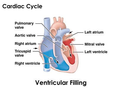
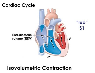
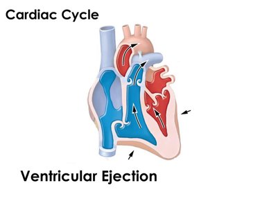
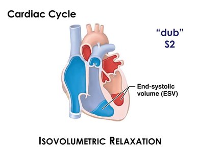
The cardiac cycle consists of sequential phases: ventricular filling (diastole), isovolumetric contraction (systole), ventricular ejection, and isovolumetric relaxation. Each phase is characterized by specific valve states and pressure changes.
Key Point 1: Ventricular filling occurs when AV valves are open and semilunar valves are closed.
Key Point 2: Isovolumetric contraction closes all valves, producing the S1 heart sound ("lub").
Key Point 3: Ventricular ejection opens semilunar valves, allowing blood to exit the heart.
Key Point 4: Isovolumetric relaxation closes all valves, producing the S2 heart sound ("dub").
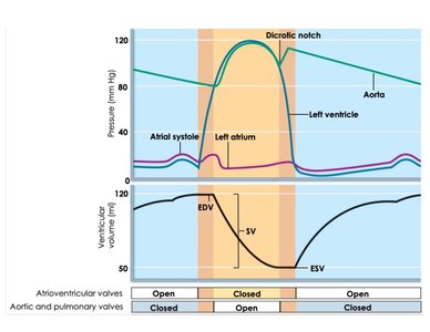
Formula: Stroke Volume (SV) = End Diastolic Volume (EDV) - End Systolic Volume (ESV)
Pressure and Volume Changes During the Cardiac Cycle
Pressure changes in the heart chambers and major vessels dictate valve states and influence ventricular volume. Blood flows down pressure gradients, and volume changes occur only when valves are open.
Key Point 1: The greatest ventricular volume occurs at the end of diastole (EDV).
Key Point 2: The smallest ventricular volume occurs at the end of systole (ESV).
Formula: Cardiac Output (CO) = Heart Rate (HR) × Stroke Volume (SV)
Summary Table: Circulatory System Types
Organism | System Type | Heart Chambers | Key Features |
|---|---|---|---|
Fish | Single Circulation | 2 (1 atrium, 1 ventricle) | Lower pressure, limited oxygen delivery |
Amphibian | Double Circulation | 3 (2 atria, 1 ventricle) | Partial separation of oxygenated/deoxygenated blood |
Mammal | Double Circulation | 4 (2 atria, 2 ventricles) | Complete separation, high efficiency |
Additional info: These notes expand upon the original slides by providing definitions, examples, and formulas relevant to General Biology students, ensuring a comprehensive understanding of circulatory system structure and function.
