 Indietro
Core Competencies and Prokaryote Diversity: General Biology Lab 1 Study Guide
Guida di studio - Note intelligenti
 Appunti personalizzati basati sui tuoi materiali, ampliati con definizioni chiave, esempi e contesto.
Lab 1: Prokaryotes
Domains of Life
Classification of Life Forms
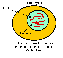
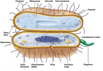
All living organisms are classified into three domains: Archaea, Bacteria, and Eukaryotes. Prokaryotes include Archaea and Bacteria, which lack a nucleus and membrane-bound organelles.
Archaea: Unique prokaryotes often found in extreme environments.
Bacteria: Diverse prokaryotes found in many habitats.
Eukaryotes: Organisms with a nucleus and complex organelles.
Domain Archaea
Characteristics and Examples
Archaea are prokaryotes that share some similarities with eukaryotes and are often adapted to extreme environments. They reproduce by binary fission and possess unique membrane lipids.
Methanococcus: Example of an archaeon, more closely related to eukaryotes than bacteria.
Halobacterium: Not a bacterium, but an archaeon. May be rod or cocci shaped, pink/red due to carotenoid pigments, motile, and UV-resistant.
Extremophiles: Many archaea thrive in extreme conditions such as high salinity, temperature, or acidity.
Domain Bacteria
Heterotrophic and Autotrophic Bacteria
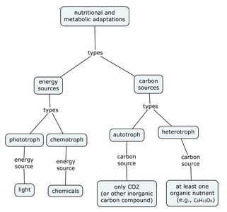
Bacteria are highly diverse and can be classified based on their nutritional and metabolic adaptations. They may be heterotrophic (consume organic matter) or autotrophic (produce their own food).
Heterotrophic bacteria: Obtain energy and carbon from organic compounds.
Photoautotrophic bacteria: Use light energy to synthesize organic compounds from CO2.
Chemoautotrophs: Use inorganic chemicals for energy; no multicellular chemoautotrophs exist.
Bacterial Shapes and Classification
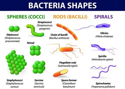
Bacteria are classified by their shapes: cocci (spheres), bacilli (rods), and spirilla (spirals). These shapes are important for identification and understanding bacterial function.
Cocci: Spherical bacteria
Bacilli: Rod-shaped bacteria
Spirilla: Spiral-shaped bacteria
Gram Staining
Principles and Procedure
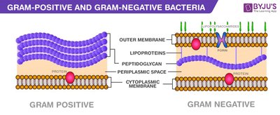
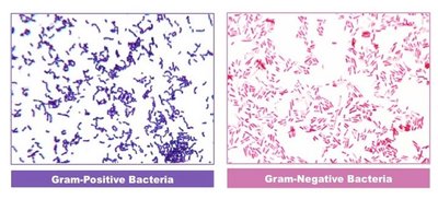
Gram staining is a fundamental technique for classifying bacteria based on cell wall structure. Gram-positive bacteria retain crystal violet stain and appear purple, while Gram-negative bacteria do not and appear pink.
Gram-positive: Thick peptidoglycan layer, stains purple.
Gram-negative: Thin peptidoglycan layer, stains pink, more resistant to antibiotics.
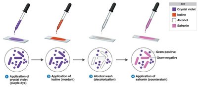
Procedure: Application of crystal violet, iodine, alcohol wash, and safranin.
Photoautotrophic Bacteria: Cyanobacteria
Role in Earth's History and Ecology
Cyanobacteria are photoautotrophic bacteria that invented oxygenic photosynthesis, transforming Earth's atmosphere and enabling the evolution of aerobic life. They are among the oldest known fossils.
Oscillatoria: Example of cyanobacteria, forms filaments.
Anabaena: Forms mutualistic relationships with plants (e.g., Azolla), contains heterocysts for nitrogen fixation.
Ecological impact: Cyanobacteria contribute to the nitrogen cycle and primary production.
Nitrogen Cycle and Bacterial Role
Biological Nitrogen Fixation
Bacteria play a crucial role in the nitrogen cycle, converting atmospheric nitrogen (N2) into forms usable by plants. This process is essential for ecosystem productivity and agriculture.
Nitrogen fixation: Conversion of N2 to ammonia by bacteria (e.g., Anabaena).
Nitrification: Conversion of ammonia to nitrite and nitrate.
Denitrification: Conversion of nitrate back to atmospheric N2.
Comparison of Key Bacterial Species
Species | Shape (Individual) | Organization | Gram -/+ | Human Impact |
|---|---|---|---|---|
Klebsiella pneumoniae | Bacillus | Chains | Negative | Pneumonia, hospital infections |
Neisseria meningitidis | Coccus | Pairs | Negative | Meningitis |
Pseudomonas aeruginosa | Bacillus | Single | Negative | Opportunistic infections |
Bacillus anthracis | Bacillus | Chains | Positive | Anthrax |
Staphylococcus epidermidis | Coccus | Clusters | Positive | Skin infections |
