 Indietro
Deuterostomes and the Evolution of Vertebrates: Structure, Diversity, and Key Innovations
Guida di studio - Note intelligenti
 Appunti personalizzati basati sui tuoi materiali, ampliati con definizioni chiave, esempi e contesto.
Deuterostome Animals
Overview of Deuterostomes
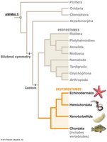
Deuterostomes are a major lineage of animals characterized by unique embryonic development and include some of the largest and most morphologically complex animals. Their development features radial cleavage, formation of the anus from the blastopore, enterocoelous coelom formation, and indeterminate fate of early blastomeres.
Radial cleavage: Cells divide parallel or perpendicular to the axis, resulting in symmetrical arrangement.
Blastopore becomes anus: The first opening in embryonic development forms the anus.
Enterocoelous: Coelom forms from mesodermal pouches.
Indeterminate cleavage: Early cells retain the ability to develop into a complete organism.
Deuterostome Phyla
Four phyla are recognized within deuterostomes, with Echinodermata and Chordata being the most studied in general biology.
Echinodermata: Sea stars, sea urchins, and relatives.
Hemichordata: Acorn worms.
Xenoturbellida: Simple, wormlike species.
Chordata: Includes vertebrates and invertebrate chordates.
Echinodermata
Defining Characteristics
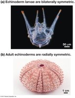
Echinoderms are exclusively marine and exhibit unique features such as pentaradial symmetry in adults, a calcium carbonate endoskeleton, and a water vascular system that forms a hydrostatic skeleton.
Bilateral larvae, pentaradial adults: Larvae are bilaterally symmetric, adults are radially symmetric.
Endoskeleton: Made of calcium carbonate plates.
Water vascular system: Network of fluid-filled canals used for movement and feeding.
Tube feet: Extensions used for locomotion and food capture.
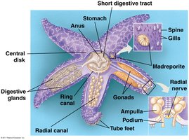
Echinoderm Life Styles
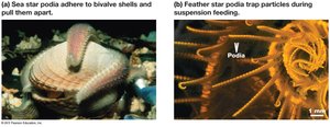
Echinoderms display diverse feeding strategies, including predation, suspension feeding, deposit feeding, and herbivory. Their podia (tube feet) are central to these behaviors.
Predatory species: Use tube feet to open prey.
Suspension feeders: Capture particles from water.
Deposit feeders: Consume organic material from substrate.
Herbivores: Feed on algae.
Echinoderm Diversity
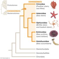
There are five major living echinoderm lineages:
Crinoidea: Feather stars and sea lilies (suspension feeders).
Asteroidea: Sea stars (predators and scavengers).
Ophiuroidea: Brittle stars and basket stars (suspension and deposit feeders).
Echinoidea: Sea urchins and sand dollars (herbivores and suspension feeders).
Holothuroidea: Sea cucumbers (suspension and deposit feeders).
Chordata
Defining Characteristics
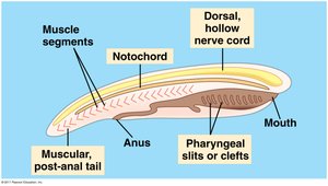
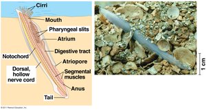
Chordates are defined by four key features present at some stage of development:
Pharyngeal slits: Openings in the pharynx used for feeding or gas exchange.
Dorsal hollow nerve cord: Runs along the back, forms the central nervous system.
Notochord: Flexible, supportive rod beneath the nerve cord.
Muscular post-anal tail: Extends beyond the anus, aids in locomotion.
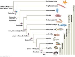
Chordate Phylogeny
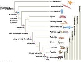
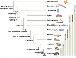
Chordates are divided into three major lineages:
Cephalochordates: Lancelets, small marine suspension feeders.
Urochordates: Tunicates, marine animals with a tunic and siphons.
Craniates (Vertebrates): Animals with a skull and often a backbone.
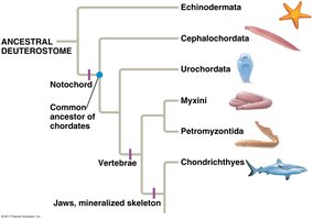
Cephalochordates
Cephalochordates, or lancelets, are small, mobile marine suspension feeders with external fertilization.
Dioecious: Separate sexes.
External fertilization: Eggs and sperm released into water.
Urochordates
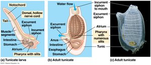
Urochordates, including tunicates and salps, are marine animals that retain only pharyngeal slits as adults and possess a tunic, U-shaped gut, and two siphons.
Exoskeletal tunic: Protective covering.
U-shaped gut: Digestive system shape.
Sexual and asexual reproduction: Some species reproduce both ways.
Craniates/Vertebrates
Craniates, or vertebrates, possess a skull, brain, sensory organs, higher metabolism, complex musculature, heart with chambers, erythrocytes with hemoglobin, and kidneys.
Spinal column: Cartilage or bone structure supporting the body.
Cranium: Protective skull for the brain.
Fin rays: Present in aquatic forms.
Large brain: Divided into forebrain, midbrain, and hindbrain.
Neural Crest Cells
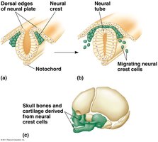
Neural crest cells are a craniate synapomorphy, forming near the dorsal margins of the closing neural tube and giving rise to craniofacial bones, melanocytes, smooth muscles, adrenal medulla, and parts of the nervous system.
Myxini (Hagfish)
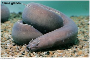
Hagfish are the most basal group of craniates, marine scavengers with a cartilaginous skull, axial rod of cartilage, and lacking jaws and vertebrae.
Slime glands: Defense mechanism.
Small brain, eyes, ears: Simple sensory organs.
Petromyzontida (Lampreys)
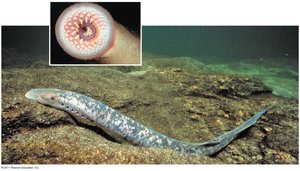
Lampreys are jawless vertebrates, ectoparasites of fish, and lack true vertebrae.
Fresh and salt water: Diverse habitats.
Jawless: Cyclostome group.
Key Vertebrate Innovations
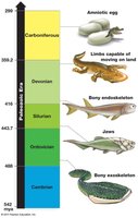
Major evolutionary innovations in vertebrates include the bony exoskeleton, jaws, bony endoskeleton, limbs capable of moving on land, and the amniotic egg.
Bony exoskeleton: First appeared ~480 mya.
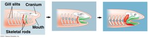
Jaws: Evolved from gill arch cartilage.
Bony endoskeleton: Internal support.
Limbs: Adapted for terrestrial movement.
Amniotic egg: Enabled reproduction independent of water.
Jaw Evolution
Jaws evolved from the anterior-most gill arch cartilage, a structure derived from neural crest cells. Gnathostomes (jawed vertebrates) are characterized by enlarged forebrains and lateral lines in aquatic forms.
Vertebrate Diversity
Major Groups of Vertebrates
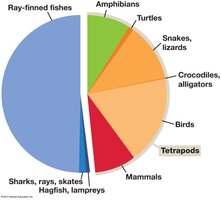
Vertebrates are classified into four major groups: jawless fishes (Myxini and Petromyzontida), cartilaginous fishes (Chondrichthyes), bony fishes (Osteichthyes), and tetrapods (amphibians, reptiles, birds, mammals).
Chondrichthyes: Cartilaginous skeleton, paired fins, internal fertilization.
Osteichthyes: Bony skeleton, operculum, swim bladder.
Tetrapods: Four limbs, adapted for land.
Osteichthyes: Bony Fishes
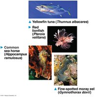
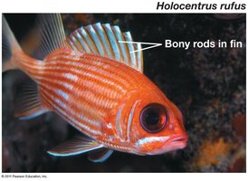
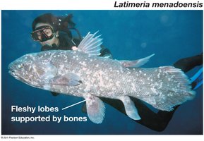
Bony fishes are divided into Actinopterygii (ray-finned fishes) and Sarcopterygii (lobe-finned fishes, including tetrapods). They possess a bony endoskeleton, operculum, lateral line system, and often a swim bladder.
Actinopterygii: Fin rays, swim bladder, diverse habitats.
Sarcopterygii: Muscular fins supported by bones, cosmine scales.
Fin-Limb Transition
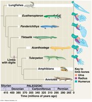
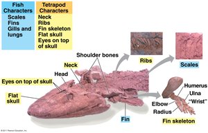
The transition from fins to limbs was a major event in vertebrate evolution, linking modern lungfish to tetrapods. Fossils such as Tiktaalik show both fish and tetrapod characteristics, supporting the hypothesis that mutation and natural selection gradually transformed fins into limbs.
Tetrapod adaptations: Four limbs with digits, neck, pelvic girdle fused to backbone, absence of gills, ears for airborne sounds.
Amniotes and Mammals
Amniotic Egg and Extraembryonic Membranes
The amniotic egg is a key adaptation for terrestrial life, providing a protective shell, water, and food supply, and additional membranes for support, gas exchange, waste isolation, and nutrient provision.
Amnion: Protective membrane.
Yolk sac: Nutrient supply.
Allantois: Waste storage.
Chorion: Gas exchange.
Mammalia
Mammals are the only living synapsids, distinguished by insulating hair or fur, three ear bones, mammary glands, enucleated red blood cells, seven cervical vertebrae, and a diaphragm. They are divided into monotremes, marsupials, and placentals.
Monotremes: Egg-laying, cloaca, low metabolic rates.
Marsupials: Young born early, nurtured in marsupium.
Placentals: Young born later, more developed.
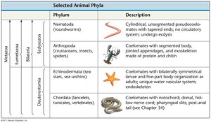
Summary Table: Deuterostome Phyla
Phylum | Description |
|---|---|
Echinodermata | Coelomates with bilaterally symmetrical larvae and five-part body organization as adults; unique water vascular system; endoskeleton |
Chordata | Coelomates with notochord; dorsal, hollow nerve cord; pharyngeal slits; post-anal tail |
Additional info: Table adapted from image_2, focusing on deuterostome phyla relevant to General Biology.
