 Indietro
Development of the Cardiovascular System: Embryonic Origins and Early Morphogenesis
Guida di studio - Note intelligenti
 Appunti personalizzati basati sui tuoi materiali, ampliati con definizioni chiave, esempi e contesto.
Cardiovascular System Development
Vasculogenesis and Angiogenesis
Vasculogenesis and angiogenesis are the processes by which the embryonic blood vessels and cardiovascular system begin to form. These processes start at the beginning of the third week of embryonic development, primarily in the yolk sac (vitellus sac), connecting stalk, and chorion.
Vasculogenesis: Formation of new blood vessels from mesodermal cells, which differentiate into angioblasts and hemangioblasts.
Angiogenesis: Growth of new blood vessels from pre-existing vessels.
Blood Islands: Clusters of mesodermal cells that give rise to both blood cells and endothelial cells.
Hematopoietic Stem Cells: Persistent stem cells develop near the mesonephric kidney, colonize the liver (hematopoietic organ between 2nd and 7th months), and later settle in the bone marrow.
Example: The liver acts as the primary site of blood cell formation during mid-gestation, before the bone marrow takes over.
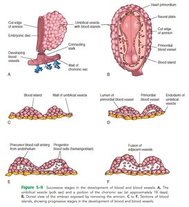
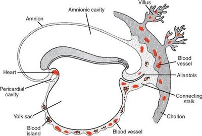
Early Heart Development
The first sign of heart development is the appearance of angioblastic cords in the cardiogenic region during the third week. Mesodermal cells migrate bilaterally along the primitive streak to form the first heart field, which will give rise to the heart.
Cardiogenic Area: Located anterior to the neural plate and buccopharyngeal membrane.
Cardiac Myoblasts: Mesodermal cells differentiate into cardiac myoblasts under the influence of the endoderm.
Heart Tube Formation: Cell communities merge to form a horseshoe-shaped tube, with an inner endothelial layer and outer myoblast layer.
Dorsal Aorta: Blood islands near the midline form paired longitudinal veins known as the dorsal aorta.
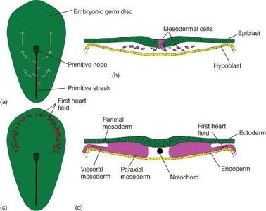
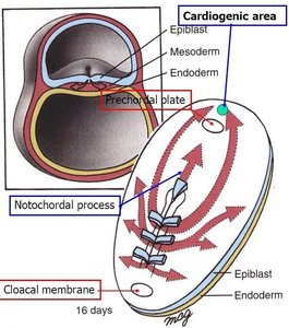
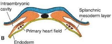
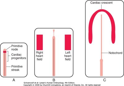
Positioning and Folding of the Heart
As the embryo undergoes cranio-caudal folding, the heart tube is repositioned ventrally and caudally. The dorsal and ventral aortae approach the cardiogenic tube, and the caudal portions fuse.
Cardiac Tube: Brought ventral to the dorsal aortae during folding.
Fusion: Caudal portions of the dorsal aortae and cardiac tube fuse, forming the primitive heart structure.
Bulbus Cordis, Ventricle, Atrium: Early heart regions begin to differentiate.
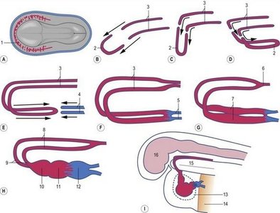
Formation of the Heart Tube and Layers
By 26 days, the heart is an uninterrupted tube lined with endothelium and surrounded by myocardium. The mesoderm adjacent to the endocardial tubes thickens to form the epimyocardial mantle, separated by cardiac jelly.
Heart Tube Layers:
Endocardium: Inner endothelial layer
Myocardium: Middle muscular layer
Epicardium: Outer layer
Cardiac Jelly: Extracellular matrix separating endothelium and myocardium, invaded by endothelial cells.
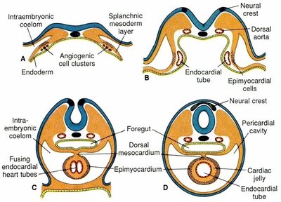
Primordial Cardiovascular System and Circulation
The heart tube fuses with blood vessels in the embryo, connecting stalk, chorion, and vitellus sac to form the primordial cardiovascular system. Heartbeat and primitive blood circulation begin at the end of the third week.
Primitive Circulation: Union of intra-embryonic and extra-embryonic vessels activates the heart.
Heartbeat: Begins on the 22nd-23rd day.
Embryonic Veins and Arteries
By the fourth week, the embryo has three pairs of veins and several arteries.
Veins:
Vitellin veins (omphalomesenteric veins)
Umbilical veins
Cardinal veins
Arteries:
Pharyngeal arch arteries
Intersegmental arteries
Vitellin arteries
Umbilical arteries
Heart Tube Growth and Bending
The heart tube grows into the pericardial cavity, initially connected by the dorsal mesocardium. The tube begins to bend on the 23rd day, forming the cardiac ring by the 28th day.
Bending: Cephalic part curves ventrally and caudally to the right; caudal atrial part curves dorsocranially to the left.
Cardiac Ring: Completed by the 28th day.
Atrioventricular Canal and Early Heart Chambers
The atrioventricular junction remains narrow, connecting the atrium to the early ventricle and forming the atrioventricular canal. The bulbus cordis forms the trabeculated part of the right ventricle, and the truncus arteriosus forms the initial part of the aorta and pulmonary artery.
Conus Cordis: Connects external flow paths of both ventricles.
Bulboventricular Sulcus: External junction between ventricle and bulbus cordis.
Primary Interventricular Foramen: Connection spot between bulbus and ventricle.
Sinus Venosus: Venous end of the heart tube.
Sinus Venosus and Venous Return
By the middle of the fourth week, the sinus venosus consists of a small transverse piece and right and left sinus horns. Each horn receives blood from three pairs of veins: vitelline, umbilical, and cardinal veins.
Vitelline Veins: Carry O2-poor blood from the yolk sac.
Umbilical Veins: Carry O2-rich blood from the chorionic villi.
Cardinal Veins: Drain the embryo body, carrying O2-poor blood.
Venous Shunt: Left-to-right shunt changes the entrance to the sinus, making the right horn dominant.
Transformation of Embryonic Veins
As development progresses, the left umbilical and left vitelline veins occlude, and the right sinus horn becomes the main passage to the atrium. The left sinus horn forms the oblique vein of the left atrium and the coronary sinus.
Cardinal Veins: Main venous drainage system; anterior and posterior cardinal veins merge and drain into the sinus venosus.
Vitellin Veins: Form hepatic sinusoids, hepatic vein, portal vein, and part of the inferior vena cava.
Umbilical Veins: Carry oxygen-rich blood from placenta; right umbilical vein degenerates, left persists.
Ductus Venosus: Shunt in the liver connecting umbilical vein to inferior vena cava, bypassing the liver.
Postnatal Changes: Umbilical vein becomes ligamentum teres; ductus venosus becomes ligamentum venosum.
Arterial System Development
The bulbus cordis enlarges and the truncus arteriosus divides to form the ascending aorta and pulmonary tract. The aortic arches develop in the neck region and differentiate into new vessels between the 6th and 8th weeks.
Aortic Arches: Originate from arcus aorticus and terminate in the dorsal aorta.
Intersegmental Arteries: Branches of the dorsal aorta that supply somites and their derivatives.
Vitellin Arteries: Supply the yolk sac and later form persistent arteries in the dorsal mesentery.
Umbilical Arteries: Double ventral branches of the dorsal aorta, heading to the placenta.
Postnatal Changes: Proximal umbilical arteries become internal iliac and superior vesical arteries; distal parts form medial umbilical ligaments.
Fetal Circulation
Fetal circulation is characterized by unique shunts that bypass certain organs to optimize oxygen delivery.
Umbilical Vein: Delivers oxygen-rich blood from placenta.
Ductus Venosus: Bypasses liver, connecting umbilical vein to inferior vena cava.
Foramen Ovale: Allows blood to flow from right atrium to left atrium, bypassing the lungs.
Ductus Arteriosus: Connects pulmonary artery to aorta, allowing blood to bypass the lungs.
Blood Distribution: 40-50% of blood in descending aorta goes to placenta via umbilical arteries; remainder supplies organs and lower body.
Example: After birth, the foramen ovale closes, and the ductus arteriosus becomes the ligamentum arteriosum.
Summary Table: Key Embryonic Veins and Their Fate
Vein | Origin | Fate |
|---|---|---|
Vitellin Vein | Yolk sac | Hepatic vein, portal vein, part of IVC |
Umbilical Vein | Placenta | Ligamentum teres (postnatal) |
Cardinal Vein | Embryo body | Vena cava, brachiocephalic veins |
Additional info: The notes have been expanded to include definitions, examples, and postnatal changes for clarity and completeness.
