Indietro
DNA Repair Mechanisms and Protein Structure: Study Notes for General Biology
Guida di studio - Note intelligenti
 Appunti personalizzati basati sui tuoi materiali, ampliati con definizioni chiave, esempi e contesto.
Repairing Mistakes and DNA Damage
Accuracy of DNA Replication
DNA replication is a highly accurate process, with an average error rate of less than one mistake per billion bases. This accuracy is primarily due to the selectivity of DNA polymerase, which matches complementary bases correctly. However, DNA polymerase can still insert an incorrect base approximately once every 100,000 bases. To maintain genetic fidelity, cells possess repair enzymes that remove and correct defective bases, especially if mistakes remain after synthesis or if DNA is damaged by external factors.
Proofreading by DNA Polymerase
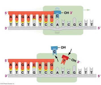
DNA polymerase has a proofreading function that checks the match between paired bases during DNA synthesis. If a mismatch is detected, the enzyme pauses and removes the mismatched base using its ε (epsilon) subunit, which acts as an exonuclease. This proofreading process reduces the error rate to about 1 × 10⁻⁷.
Mismatch Repair Mechanism
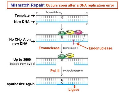
If DNA polymerase leaves a mismatched pair behind, a set of enzymes known as mismatch repair enzymes corrects the error after DNA synthesis is complete. These enzymes recognize the mismatched pair, remove a section of the newly synthesized strand containing the incorrect base, and fill in the correct bases. This process is essential for maintaining genetic stability.
Repairing Damaged DNA
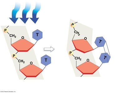
DNA can be damaged by chemicals and radiation, such as ultraviolet (UV) light, which can cause thymine dimers to form. Thymine dimers create a kink in the DNA strand, disrupting normal base pairing and replication.
Nucleotide Excision Repair
The nucleotide excision repair system recognizes and removes damaged sections of DNA, such as those containing thymine dimers. Enzymes excise the damaged single-stranded DNA, and the complementary strand serves as a template for resynthesis of the correct sequence.
Genetic Disorders and DNA Repair
Xeroderma pigmentosum (XP) is a rare autosomal recessive disease caused by mutations in nucleotide excision repair genes. Individuals with XP cannot repair DNA damaged by UV radiation, leading to the development of skin lesions and a high risk of skin cancer.
DNA Repair Genes and Cancer
Defects in DNA repair genes are frequently associated with cancer. If mutations in genes involved in the cell cycle go unrepaired, cells may grow uncontrollably, forming tumors. An elevated mutation rate due to defective DNA repair increases the likelihood of cancer-causing mutations.
Protein Structure and Function
Functions of Proteins
Proteins are essential macromolecules involved in nearly all cellular activities. Their functions include:
Catalysis: Enzymes speed up chemical reactions.
Defense: Antibodies and complement proteins attack pathogens.
Movement: Motor and contractile proteins move cells or molecules within cells.
Signaling: Proteins convey signals between cells.
Structure: Structural proteins define cell shape and form body structures.
Transport: Transport proteins carry materials; membrane proteins control molecular movement into and out of cells.
Protein Composition
Proteins are polymers made from amino acids linked by peptide bonds. There are 20 different amino acid monomers, and protein diversity arises from different arrangements of these amino acids.
Structure of Amino Acids
All amino acids share a central carbon atom bonded to:
A hydrogen atom (H)
An amino group (NH2)
A carboxyl group (COOH)
A variable side chain (R-group)
At physiological pH (7), the amino group is ionized to NH3+ and the carboxyl group to COO–, increasing solubility and reactivity.
Classification of Amino Acids by R Group
Amino acids are classified based on the properties of their R-groups:
Polar Charged: R-group has a positive or negative charge (acidic or basic, hydrophilic).
Polar Uncharged: R-group contains an oxygen atom but no charge (hydrophilic).
Nonpolar: R-group lacks charge and oxygen (hydrophobic).
R-groups differ in size, shape, reactivity, and interactions with water, influencing protein structure and function.
Polymerization and Hydrolysis
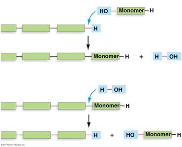
Polymerization of amino acids into proteins is a nonspontaneous process requiring energy. It occurs via condensation (dehydration) reactions, which release water. The reverse process, hydrolysis, breaks polymers apart by adding water.
Peptide Bond Formation
A peptide bond forms between the carboxyl group of one amino acid and the amino group of another via a condensation reaction. Chains of amino acids are called polypeptides. Polypeptides with fewer than 50 amino acids are called oligopeptides (peptides), while those with more than 50 are considered proteins.

Polypeptide Characteristics
The peptide-bonded backbone of a polypeptide has three key characteristics:
R-group orientation: Side chains can interact with each other or water.
Directionality: The N-terminus (free amino group) is on the left; the C-terminus (free carboxyl group) is on the right.
Flexibility: Single bonds on either side of the peptide bond can rotate, making the structure flexible.
Levels of Protein Structure
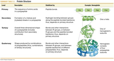
Primary Structure
The primary structure of a protein is its unique sequence of amino acids. This sequence determines all higher levels of structure and ultimately the protein's function. Even a single amino acid change can radically alter protein function, as seen in diseases like sickle cell anemia.
Secondary Structure
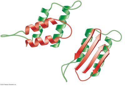
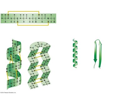
The secondary structure is formed by hydrogen bonds between the carbonyl group of one amino acid and the amino group of another. Common secondary structures include α-helices and β-pleated sheets. The propensity for forming these structures depends on the primary sequence.
Tertiary Structure
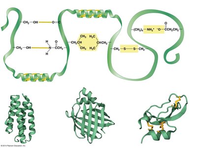
The tertiary structure results from interactions between R-groups or between R-groups and the peptide backbone. These interactions include hydrogen bonds, hydrophobic interactions, van der Waals interactions, covalent disulfide bonds, and ionic bonds. The tertiary structure gives the protein its three-dimensional shape.
Quaternary Structure
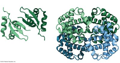
Some proteins consist of multiple polypeptide subunits. The quaternary structure is the arrangement of these subunits into a single functional complex. Examples include hemoglobin, which is composed of four subunits.
Summary Table: Protein Structure
Level | Description | Stabilized by | Example |
|---|---|---|---|
Primary | Sequence of amino acids | Peptide bonds | Hemoglobin subunit |
Secondary | α-helices and β-sheets | Hydrogen bonds | One α-helix |
Tertiary | Three-dimensional shape | R-group interactions | One hemoglobin subunit |
Quaternary | Multiple polypeptides | Subunit interactions | Hemoglobin complex |
Protein Folding and Function
Folding and Stability
Protein folding is often spontaneous due to hydrogen bonds and van der Waals interactions, which stabilize the folded molecule. A denatured (unfolded) protein cannot function normally. In some cases, molecular chaperones assist in proper folding, especially under stress conditions (e.g., heat shock proteins).
Regulation of Protein Folding
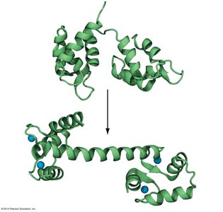
Protein folding is regulated because function depends on shape. Proteins may exist in inactive, disordered forms and fold into active conformations when needed. This regulation ensures proteins are active only when required.
Prions and Protein Misfolding
Prions are infectious, misfolded forms of normal proteins. Although their amino acid sequence is unchanged, their altered shape can induce normal proteins to adopt the misfolded, pathogenic form. Prions are associated with neurodegenerative diseases.